
21.1 : Anatomie des systèmes lymphatique et immunitaire
- Page ID
- 195484

Objectifs d'apprentissage
- Décrire la structure et la fonction du tissu lymphatique (liquide lymphatique, vaisseaux, canaux et organes)
- Décrire la structure et la fonction des organes lymphatiques primaires et secondaires
- Discutez des cellules du système immunitaire, de leur fonctionnement et de leur relation avec le système lymphatique
Le système immunitaire est un ensemble complexe de cellules et d'organes qui détruit ou neutralise les agents pathogènes qui, autrement, provoqueraient des maladies ou la mort. Pour la plupart des gens, le système lymphatique est associé au système immunitaire à tel point que les deux systèmes sont pratiquement impossibles à distinguer. Le système lymphatique est l'ensemble des vaisseaux, des cellules et des organes qui transportent l'excès de liquide vers la circulation sanguine et filtrent les agents pathogènes présents dans le sang. Le gonflement des ganglions lymphatiques lors d'une infection et le transport des lymphocytes par les vaisseaux lymphatiques ne sont que deux exemples des nombreuses connexions entre ces systèmes organiques critiques.
Fonctions du système lymphatique
L'une des principales fonctions du système lymphatique est de drainer les fluides corporels et de les renvoyer dans la circulation sanguine. La pression artérielle provoque une fuite de liquide par les capillaires, ce qui entraîne l'accumulation de liquide dans l'espace interstitiel, c'est-à-dire les espaces entre les cellules individuelles des tissus. Chez l'homme, 20 litres de plasma sont libérés chaque jour dans l'espace interstitiel des tissus grâce à la filtration capillaire. Une fois que ce filtrat sort de la circulation sanguine et se retrouve dans les espaces tissulaires, on parle de liquide interstitiel. De ce montant, 17 litres sont réabsorbés directement par les vaisseaux sanguins. Mais qu'advient-il des trois litres restants ? C'est là que le système lymphatique entre en jeu. Il draine l'excès de liquide et le renvoie dans la circulation sanguine par une série de vaisseaux, de troncs et de conduits. La lymphe est le terme utilisé pour décrire le liquide interstitiel une fois qu'il a pénétré dans le système lymphatique. Lorsque le système lymphatique est endommagé d'une manière ou d'une autre, par exemple en étant obstrué par des cellules cancéreuses ou détruit par une blessure, le liquide interstitiel riche en protéines s'accumule (parfois « remonte » des vaisseaux lymphatiques) dans les espaces tissulaires. Cette accumulation inappropriée de liquide appelée lymphœdème peut entraîner de graves conséquences médicales.
Au fur et à mesure de l'évolution du système immunitaire des vertébrés, le réseau de vaisseaux lymphatiques est devenu un moyen pratique de transport des cellules du système immunitaire. De plus, le transport des lipides alimentaires et des vitamines liposolubles absorbés par l'intestin utilise ce système.
Les cellules du système immunitaire utilisent non seulement les vaisseaux lymphatiques pour passer des espaces interstitiels à la circulation, mais elles utilisent également les ganglions lymphatiques comme principaux stades du développement de réponses immunitaires critiques. Un ganglion lymphatique est l'un des petits organes en forme de haricot situés dans tout le système lymphatique.

Visitez ce site Web pour un aperçu du système lymphatique. Quels sont les trois principaux composants du système lymphatique ?
Structure du système lymphatique
Les vaisseaux lymphatiques prennent d'abord la forme de capillaires à extrémité ouverte, qui alimentent des vaisseaux lymphatiques de plus en plus grands et finissent par se déverser dans la circulation sanguine par une série de canaux. En cours de route, la lymphe traverse les ganglions lymphatiques, que l'on trouve généralement près de l'aine, des aisselles, du cou, de la poitrine et de l'abdomen. Les humains ont environ 500 à 600 ganglions lymphatiques dans tout le corps (Figure\(\PageIndex{1}\)).
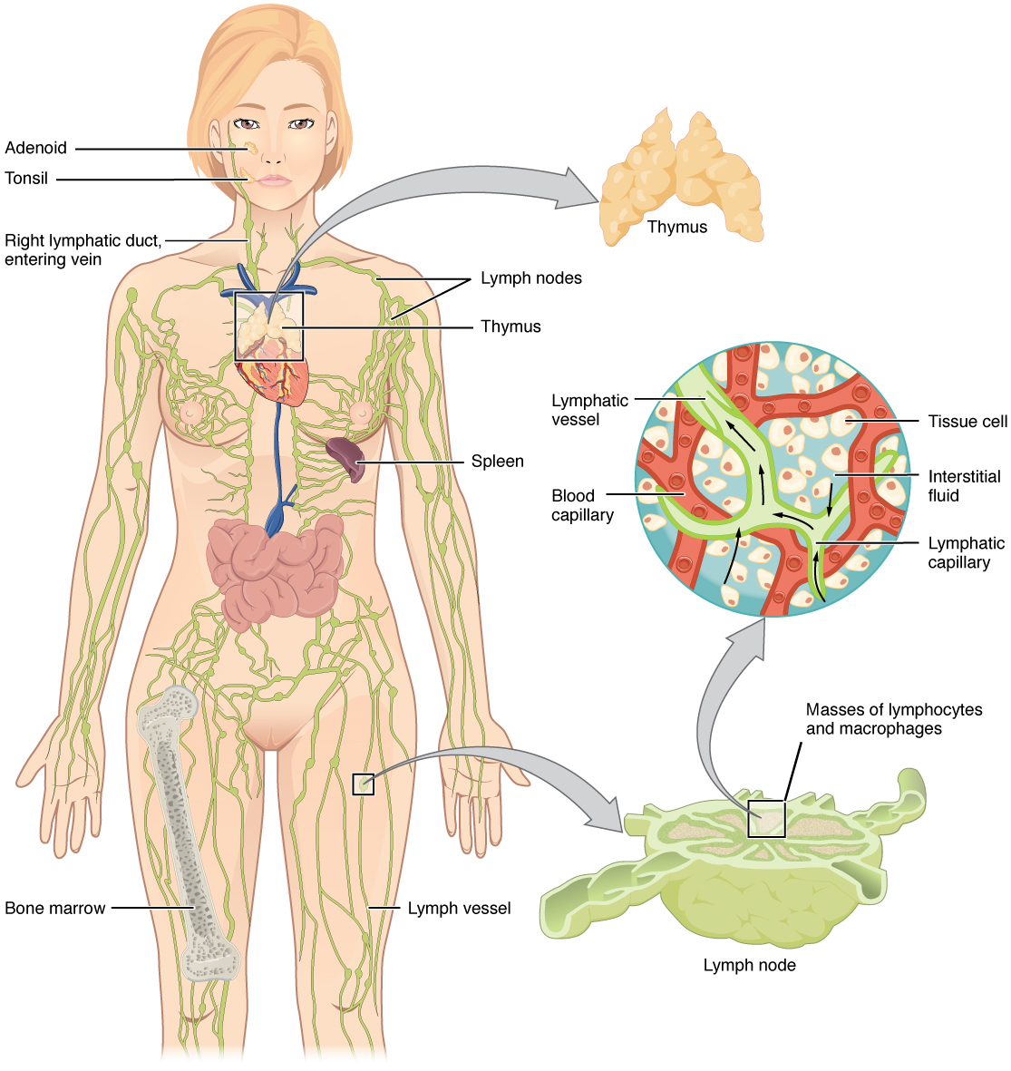
A major distinction between the lymphatic and cardiovascular systems in humans is that lymph is not actively pumped by the heart, but is forced through the vessels by the movements of the body, the contraction of skeletal muscles during body movements, and breathing. One-way valves (semi-lunar valves) in lymphatic vessels keep the lymph moving toward the heart. Lymph flows from the lymphatic capillaries, through lymphatic vessels, and then is dumped into the circulatory system via the lymphatic ducts located at the junction of the jugular and subclavian veins in the neck.
Lymphatic Capillaries
Lymphatic capillaries, also called the terminal lymphatics, are vessels where interstitial fluid enters the lymphatic system to become lymph fluid. Located in almost every tissue in the body, these vessels are interlaced among the arterioles and venules of the circulatory system in the soft connective tissues of the body (Figure \(\PageIndex{2}\)). Exceptions are the central nervous system, bone marrow, bones, teeth, and the cornea of the eye, which do not contain lymph vessels.
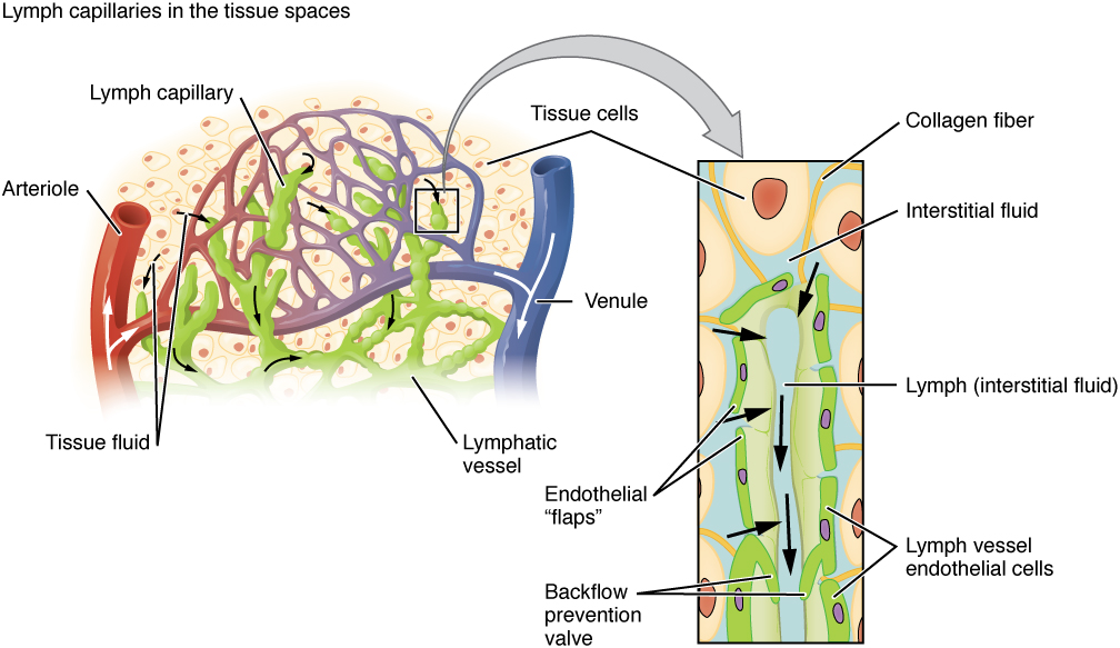
Lymphatic capillaries are formed by a one cell-thick layer of endothelial cells and represent the open end of the system, allowing interstitial fluid to flow into them via overlapping cells (see Figure \(\PageIndex{2}\)). When interstitial pressure is low, the endothelial flaps close to prevent “backflow.” As interstitial pressure increases, the spaces between the cells open up, allowing the fluid to enter. Entry of fluid into lymphatic capillaries is also enabled by the collagen filaments that anchor the capillaries to surrounding structures. As interstitial pressure increases, the filaments pull on the endothelial cell flaps, opening up them even further to allow easy entry of fluid.
In the small intestine, lymphatic capillaries called lacteals are critical for the transport of dietary lipids and lipid-soluble vitamins to the bloodstream. In the small intestine, dietary triglycerides combine with other lipids and proteins, and enter the lacteals to form a milky fluid called chyle. The chyle then travels through the lymphatic system, eventually entering the liver and then the bloodstream.
Larger Lymphatic Vessels, Trunks, and Ducts
The lymphatic capillaries empty into larger lymphatic vessels, which are similar to veins in terms of their three-tunic structure and the presence of valves. These one-way valves are located fairly close to one another, and each one causes a bulge in the lymphatic vessel, giving the vessels a beaded appearance (see Figure \(\PageIndex{2}\)).
The superficial and deep lymphatics eventually merge to form larger lymphatic vessels known as lymphatic trunks. On the right side of the body, the right sides of the head, thorax, and right upper limb drain lymph fluid into the right subclavian vein via the right lymphatic duct (Figure \(\PageIndex{3}\)). On the left side of the body, the remaining portions of the body drain into the larger thoracic duct, which drains into the left subclavian vein. The thoracic duct itself begins just beneath the diaphragm in the cisterna chyli, a sac-like chamber that receives lymph from the lower abdomen, pelvis, and lower limbs by way of the left and right lumbar trunks and the intestinal trunk.

The overall drainage system of the body is asymmetrical (see Figure \(\PageIndex{3}\)). The right lymphatic duct receives lymph from only the upper right side of the body. The lymph from the rest of the body enters the bloodstream through the thoracic duct via all the remaining lymphatic trunks. In general, lymphatic vessels of the subcutaneous tissues of the skin, that is, the superficial lymphatics, follow the same routes as veins, whereas the deep lymphatic vessels of the viscera generally follow the paths of arteries.
The Organization of Immune Function
The immune system is a collection of barriers, cells, and soluble proteins that interact and communicate with each other in extraordinarily complex ways. The modern model of immune function is organized into three phases based on the timing of their effects. The three temporal phases consist of the following:
- Barrier defenses such as the skin and mucous membranes, which act instantaneously to prevent pathogenic invasion into the body tissues
- The rapid but nonspecific innate immune response, which consists of a variety of specialized cells and soluble factors
- The slower but more specific and effective adaptive immune response, which involves many cell types and soluble factors, but is primarily controlled by white blood cells (leukocytes) known as lymphocytes, which help control immune responses
The cells of the blood, including all those involved in the immune response, arise in the bone marrow via various differentiation pathways from hematopoietic stem cells (Figure \(\PageIndex{4}\)). In contrast with embryonic stem cells, hematopoietic stem cells are present throughout adulthood and allow for the continuous differentiation of blood cells to replace those lost to age or function. These cells can be divided into three classes based on function:
- Phagocytic cells, which ingest pathogens to destroy them
- Lymphocytes, which specifically coordinate the activities of adaptive immunity
- Cells containing cytoplasmic granules, which help mediate immune responses against parasites and intracellular pathogens such as viruses

Lymphocytes: B Cells, T Cells, Plasma Cells, and Natural Killer Cells
As stated above, lymphocytes are the primary cells of adaptive immune responses (Table). The two basic types of lymphocytes, B cells and T cells, are identical morphologically with a large central nucleus surrounded by a thin layer of cytoplasm. They are distinguished from each other by their surface protein markers as well as by the molecules they secrete. While B cells mature in red bone marrow and T cells mature in the thymus, they both initially develop from bone marrow. T cells migrate from bone marrow to the thymus gland where they further mature. B cells and T cells are found in many parts of the body, circulating in the bloodstream and lymph, and residing in secondary lymphoid organs, including the spleen and lymph nodes, which will be described later in this section. The human body contains approximately 1012 lymphocytes.
B Cells
B cells are immune cells that function primarily by producing antibodies. An antibody is any of the group of proteins that binds specifically to pathogen-associated molecules known as antigens. An antigen is a chemical structure on the surface of a pathogen that binds to T or B lymphocyte antigen receptors. Once activated by binding to antigen, B cells differentiate into cells that secrete a soluble form of their surface antibodies. These activated B cells are known as plasma cells.
T Cells
The T cell, on the other hand, does not secrete antibody but performs a variety of functions in the adaptive immune response. Different T cell types have the ability to either secrete soluble factors that communicate with other cells of the adaptive immune response or destroy cells infected with intracellular pathogens. The roles of T and B lymphocytes in the adaptive immune response will be discussed further in this chapter.
Plasma Cells
Another type of lymphocyte of importance is the plasma cell. A plasma cell is a B cell that has differentiated in response to antigen binding, and has thereby gained the ability to secrete soluble antibodies. These cells differ in morphology from standard B and T cells in that they contain a large amount of cytoplasm packed with the protein-synthesizing machinery known as rough endoplasmic reticulum.
Natural Killer Cells
A fourth important lymphocyte is the natural killer cell, a participant in the innate immune response. A natural killer cell (NK) is a circulating blood cell that contains cytotoxic (cell-killing) granules in its extensive cytoplasm. It shares this mechanism with the cytotoxic T cells of the adaptive immune response. NK cells are among the body’s first lines of defense against viruses and certain types of cancer.
| Lymphocytes | |
|---|---|
| Type of lymphocyte | Primary function |
| B lymphocyte | Generates diverse antibodies |
| T lymphocyte | Secretes chemical messengers |
| Plasma cell | Secretes antibodies |
| NK cell | Destroys virally infected cells |

Visitez ce site Web pour en savoir plus sur les nombreux types de cellules du système immunitaire et leurs fonctions très spécialisées. Quel est le rôle de la cellule dendritique dans l'infection par le VIH ?
Organes lymphoïdes primaires et développement des lymphocytes
Il est essentiel de comprendre la différenciation et le développement des lymphocytes B et T pour comprendre la réponse immunitaire adaptative. C'est grâce à ce processus que le corps apprend (idéalement) à détruire uniquement les agents pathogènes et laisse ses propres cellules relativement intactes. Les principaux organes lymphoïdes sont la moelle osseuse, la rate et le thymus. Les organes lymphoïdes sont l'endroit où les lymphocytes mûrissent, prolifèrent et sont sélectionnés, ce qui leur permet d'attaquer les agents pathogènes sans endommager les cellules du corps.
Moelle osseuse
Dans l'embryon, les cellules sanguines sont fabriquées dans le sac vitellin. Au fur et à mesure du développement, cette fonction est assumée par la rate, les ganglions lymphatiques et le foie. Plus tard, la moelle osseuse assume la plupart des fonctions hématopoïétiques, bien que les derniers stades de la différenciation de certaines cellules puissent avoir lieu dans d'autres organes. La moelle osseuse rouge est un ensemble lâche de cellules où se produit l'hématopoïèse, et la moelle osseuse jaune est un site de stockage d'énergie, composé en grande partie de cellules graisseuses (Figure\(\PageIndex{5}\)). Le lymphocyte B se développe presque entièrement dans la moelle osseuse rouge, tandis que le lymphocyte T immature, appelé thymocyte, quitte la moelle osseuse et mûrit en grande partie dans le thymus.

Thymus
Le thymus est un organe bilobé situé dans l'espace situé entre le sternum et l'aorte du cœur (Figure\(\PageIndex{6}\)). Le tissu conjonctif maintient les lobes étroitement les uns contre les autres mais les sépare et forme une capsule.
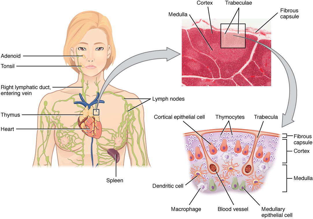

La capsule du tissu conjonctif divise ensuite le thymus en lobules via des extensions appelées trabécules. La région externe de l'organe est connue sous le nom de cortex et contient un grand nombre de thymocytes ainsi que des cellules épithéliales, des macrophages et des cellules dendritiques (deux types de cellules phagocytaires dérivées de monocytes). Le cortex est dense, de sorte qu'il colore plus intensément que le reste du thymus (voir Figure\(\PageIndex{6}\)). The medulla, where thymocytes migrate before leaving the thymus, contains a less dense collection of thymocytes, epithelial cells, and dendritic cells.
AGING AND THE...
Immune System
By the year 2050, 25 percent of the population of the United States will be 60 years of age or older. The CDC estimates that 80 percent of those 60 years and older have one or more chronic disease associated with deficiencies of the immune systems. This loss of immune function with age is called immunosenescence. To treat this growing population, medical professionals must better understand the aging process. One major cause of age-related immune deficiencies is thymic involution, the shrinking of the thymus gland that begins at birth, at a rate of about three percent tissue loss per year, and continues until 35–45 years of age, when the rate declines to about one percent loss per year for the rest of one’s life. At that pace, the total loss of thymic epithelial tissue and thymocytes would occur at about 120 years of age. Thus, this age is a theoretical limit to a healthy human lifespan.
Thymic involution has been observed in all vertebrate species that have a thymus gland. Animal studies have shown that transplanted thymic grafts between inbred strains of mice involuted according to the age of the donor and not of the recipient, implying the process is genetically programmed. There is evidence that the thymic microenvironment, so vital to the development of naïve T cells, loses thymic epithelial cells according to the decreasing expression of the FOXN1 gene with age.
It is also known that thymic involution can be altered by hormone levels. Sex hormones such as estrogen and testosterone enhance involution, and the hormonal changes in pregnant women cause a temporary thymic involution that reverses itself, when the size of the thymus and its hormone levels return to normal, usually after lactation ceases. What does all this tell us? Can we reverse immunosenescence, or at least slow it down? The potential is there for using thymic transplants from younger donors to keep thymic output of naïve T cells high. Gene therapies that target gene expression are also seen as future possibilities. The more we learn through immunosenescence research, the more opportunities there will be to develop therapies, even though these therapies will likely take decades to develop. The ultimate goal is for everyone to live and be healthy longer, but there may be limits to immortality imposed by our genes and hormones.
Secondary Lymphoid Organs and their Roles in Active Immune Responses
Lymphocytes develop and mature in the primary lymphoid organs, but they mount immune responses from the secondary lymphoid organs. A naïve lymphocyte is one that has left the primary organ and entered a secondary lymphoid organ. Naïve lymphocytes are fully functional immunologically, but have yet to encounter an antigen to respond to. In addition to circulating in the blood and lymph, lymphocytes concentrate in secondary lymphoid organs, which include the lymph nodes, spleen, and lymphoid nodules. All of these tissues have many features in common, including the following:
- The presence of lymphoid follicles, the sites of the formation of lymphocytes, with specific B cell-rich and T cell-rich areas
- An internal structure of reticular fibers with associated fixed macrophages
- Germinal centers, which are the sites of rapidly dividing B lymphocytes and plasma cells, with the exception of the spleen
- Specialized post-capillary vessels known as high endothelial venules; the cells lining these venules are thicker and more columnar than normal endothelial cells, which allow cells from the blood to directly enter these tissues
Lymph Nodes
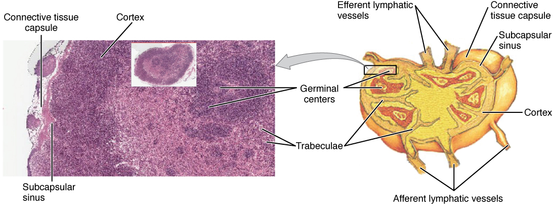
Lymph nodes function to remove debris and pathogens from the lymph, and are thus sometimes referred to as the “filters of the lymph” (Figure \(\PageIndex{7}\)). Any bacteria that infect the interstitial fluid are taken up by the lymphatic capillaries and transported to a regional lymph node. Dendritic cells and macrophages within this organ internalize and kill many of the pathogens that pass through, thereby removing them from the body. The lymph node is also the site of adaptive immune responses mediated by T cells, B cells, and accessory cells of the adaptive immune system. Like the thymus, the bean-shaped lymph nodes are surrounded by a tough capsule of connective tissue and are separated into compartments by trabeculae, the extensions of the capsule. In addition to the structure provided by the capsule and trabeculae, the structural support of the lymph node is provided by a series of reticular fibers laid down by fibroblasts.

Consultez le WebScope de l'Université du Michigan pour explorer l'échantillon de tissu plus en détail.
Les principales voies d'accès au ganglion lymphatique passent par les vaisseaux lymphatiques afférents (voir Figure\(\PageIndex{7}\)). Les cellules et le liquide lymphatique qui quittent le ganglion lymphatique peuvent le faire par un autre ensemble de vaisseaux appelés vaisseaux lymphatiques efférents. La lymphe entre dans le ganglion lymphatique par le sinus sous-capsulaire, qui est occupé par des cellules dendritiques, des macrophages et des fibres réticulaires. Dans le cortex du ganglion lymphatique se trouvent des follicules lymphoïdes, qui sont constitués de centres germinaux de lymphocytes B à division rapide entourés d'une couche de lymphocytes T et d'autres cellules accessoires. Au fur et à mesure que la lymphe traverse le ganglion, elle pénètre dans la moelle, qui est constituée de cordons médullaires de lymphocytes B et de plasmocytes, et dans les sinus médullaires où la lymphe s'accumule avant de quitter le ganglion par les vaisseaux lymphatiques efférents.
Rate
Outre les ganglions lymphatiques, la rate est un organe lymphoïde secondaire majeur (Figure\(\PageIndex{8}\)). Il mesure environ 12 cm (5 po) de long et est attaché au bord latéral de l'estomac par le ligament gastrosplénique. La rate est un organe fragile sans capsule forte et est rouge foncé en raison de sa vascularisation étendue. La rate est parfois appelée le « filtre du sang » en raison de sa vascularisation étendue et de la présence de macrophages et de cellules dendritiques qui éliminent les microbes et autres substances du sang, y compris les globules rouges mourants. La rate joue également le rôle de localisation des réponses immunitaires aux agents pathogènes transmis par le sang.

La rate est également divisée par des trabécules de tissu conjonctif et, à l'intérieur de chaque nodule splénique, se trouve une zone de pulpe rouge, composée principalement de globules rouges, et de pulpe blanche, qui ressemble aux follicules lymphoïdes des ganglions lymphatiques. En pénétrant dans la rate, l'artère splénique se divise en plusieurs artérioles (entourées de pulpe blanche) et finalement en sinusoïdes. Le sang provenant des capillaires s'accumule ensuite dans les sinus veineux et sort par la veine splénique. La pulpe rouge est constituée de fibres réticulaires auxquelles sont attachés des macrophages fixes, de macrophages libres et de toutes les autres cellules typiques du sang, y compris certains lymphocytes. La pulpe blanche entoure une artériole centrale et est constituée de centres germinaux de lymphocytes B en division entourés de lymphocytes T et de cellules accessoires, y compris des macrophages et des cellules dendritiques. Ainsi, la pulpe rouge fonctionne principalement comme un système de filtration du sang, utilisant des cellules présentant une réponse immunitaire relativement non spécifique, et la pulpe blanche est l'endroit où se produisent les réponses adaptatives des lymphocytes T et B.
Nodules lymphoïdes
Les autres tissus lymphoïdes, les nodules lymphoïdes, ont une architecture plus simple que celle de la rate et des ganglions lymphatiques en ce sens qu'ils sont constitués d'un amas dense de lymphocytes sans capsule fibreuse environnante. Ces nodules sont localisés dans les voies respiratoires et digestives, zones régulièrement exposées aux agents pathogènes de l'environnement.
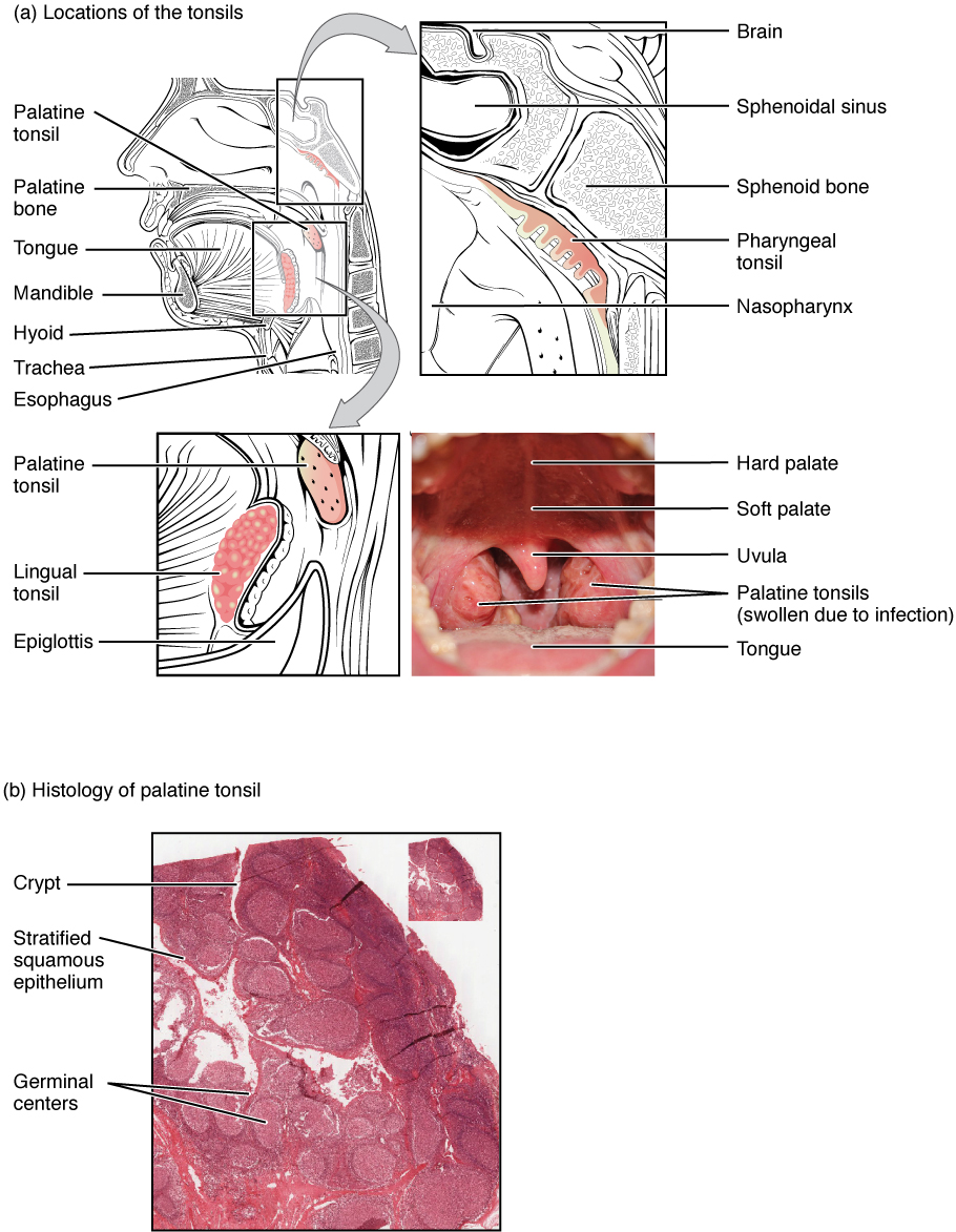
Les amygdales sont des nodules lymphoïdes situés le long de la surface interne du pharynx et jouent un rôle important dans le développement de l'immunité contre les agents pathogènes de la bouche (Figure\(\PageIndex{9}\)). L'amygdale située à l'arrière de la gorge, l'amygdale pharyngée, est parfois appelée adénoïde lorsqu'elle est enflée. Un tel gonflement indique une réponse immunitaire active à l'infection. Sur le plan histologique, les amygdales ne contiennent pas de capsule complète et la couche épithéliale pénètre profondément à l'intérieur de l'amygdale pour former des cryptes amygdaliennes. Ces structures, qui accumulent toutes sortes de matières absorbées par l'organisme lors de l'alimentation et de la respiration, « encouragent » en fait les agents pathogènes à pénétrer profondément dans les tissus amygdaliens où ils sont soumis à l'action de nombreux follicules lymphoïdes et éliminés. Cela semble être la principale fonction des amygdales : aider le corps des enfants à reconnaître, à détruire et à développer une immunité contre les agents pathogènes environnementaux courants afin qu'ils soient protégés plus tard dans leur vie. Les amygdales sont souvent enlevées chez les enfants atteints d'infections récurrentes de la gorge, en particulier celles qui touchent les amygdales palatines de chaque côté de la gorge, dont l'enflure peut gêner leur respiration et/ou leur déglutition.

Le tissu lymphoïde associé aux muqueuses (MALT) est constitué d'un agrégat de follicules lymphoïdes directement associés à l'épithélium de la muqueuse. Le MALT est constitué de structures en forme de dôme situées sous la muqueuse du tractus gastro-intestinal, des tissus mammaires, des poumons et des yeux. Les plaques de Peyer, un type de MALT présent dans l'intestin grêle, sont particulièrement importantes pour les réponses immunitaires contre les substances ingérées (Figure\(\PageIndex{10}\)). Peyer’s patches contain specialized endothelial cells called M (or microfold) cells that sample material from the intestinal lumen and transport it to nearby follicles so that adaptive immune responses to potential pathogens can be mounted.
_Nodule.jpg)
Bronchus-associated lymphoid tissue (BALT) consists of lymphoid follicular structures with an overlying epithelial layer found along the bifurcations of the bronchi, and between bronchi and arteries. They also have the typically less-organized structure of other lymphoid nodules. These tissues, in addition to the tonsils, are effective against inhaled pathogens.
Chapter Review
The lymphatic system is a series of vessels, ducts, and trunks that remove interstitial fluid from the tissues and return it the blood. The lymphatics are also used to transport dietary lipids and cells of the immune system. Cells of the immune system all come from the hematopoietic system of the bone marrow. Primary lymphoid organs, the bone marrow and thymus gland, are the locations where lymphocytes of the adaptive immune system proliferate and mature. Secondary lymphoid organs are site in which mature lymphocytes congregate to mount immune responses. Many immune system cells use the lymphatic and circulatory systems for transport throughout the body to search for and then protect against pathogens.
Interactive Link Questions
Visit this website for an overview of the lymphatic system. What are the three main components of the lymphatic system?
Answer: The three main components are the lymph vessels, the lymph nodes, and the lymph.
Visit this website to learn about the many different cell types in the immune system and their very specialized jobs. What is the role of the dendritic cell in infection by HIV?
Answer: The dendritic cell transports the virus to a lymph node.
Review Questions
Q. Which of the following cells is phagocytic?
A. plasma cell
B. macrophage
C. B cell
D. NK cell
Answer: B
Q. Which structure allows lymph from the lower right limb to enter the bloodstream?
A. thoracic duct
B. right lymphatic duct
C. right lymphatic trunk
D. left lymphatic trunk
Answer: A
Q. Which of the following cells is important in the innate immune response?
A. B cells
B. T cells
C. macrophages
D. plasma cells
Answer: C
Q. Which of the following cells would be most active in early, antiviral immune responses the first time one is exposed to pathogen?
A. macrophage
B. T cell
E. neutrophil
D. natural killer cell
Answer: D
Q. Which of the lymphoid nodules is most likely to see food antigens first?
A. tonsils
B. Peyer’s patches
C. bronchus-associated lymphoid tissue
D. mucosa-associated lymphoid tissue
Answer: A
Critical Thinking Questions
Q. Describe the flow of lymph from its origins in interstitial fluid to its emptying into the venous bloodstream.
A. The lymph enters through lymphatic capillaries, and then into larger lymphatic vessels. The lymph can only go in one direction due to valves in the vessels. The larger lymphatics merge to form trunks that enter into the blood via lymphatic ducts.
Glossary
- adaptive immune response
- relatively slow but very specific and effective immune response controlled by lymphocytes
- afferent lymphatic vessels
- lead into a lymph node
- antibody
- antigen-specific protein secreted by plasma cells; immunoglobulin
- antigen
- molecule recognized by the receptors of B and T lymphocytes
- barrier defenses
- antipathogen defenses deriving from a barrier that physically prevents pathogens from entering the body to establish an infection
- B cells
- lymphocytes that act by differentiating into an antibody-secreting plasma cell
- bone marrow
- tissue found inside bones; the site of all blood cell differentiation and maturation of B lymphocytes
- bronchus-associated lymphoid tissue (BALT)
- lymphoid nodule associated with the respiratory tract
- chyle
- lipid-rich lymph inside the lymphatic capillaries of the small intestine
- cisterna chyli
- bag-like vessel that forms the beginning of the thoracic duct
- efferent lymphatic vessels
- lead out of a lymph node
- germinal centers
- clusters of rapidly proliferating B cells found in secondary lymphoid tissues
- high endothelial venules
- vessels containing unique endothelial cells specialized to allow migration of lymphocytes from the blood to the lymph node
- immune system
- series of barriers, cells, and soluble mediators that combine to response to infections of the body with pathogenic organisms
- innate immune response
- rapid but relatively nonspecific immune response
- lymph
- fluid contained within the lymphatic system
- lymph node
- one of the bean-shaped organs found associated with the lymphatic vessels
- lymphatic capillaries
- smallest of the lymphatic vessels and the origin of lymph flow
- lymphatic system
- network of lymphatic vessels, lymph nodes, and ducts that carries lymph from the tissues and back to the bloodstream.
- lymphatic trunks
- large lymphatics that collect lymph from smaller lymphatic vessels and empties into the blood via lymphatic ducts
- lymphocytes
- white blood cells characterized by a large nucleus and small rim of cytoplasm
- lymphoid nodules
- unencapsulated patches of lymphoid tissue found throughout the body
- mucosa-associated lymphoid tissue (MALT)
- lymphoid nodule associated with the mucosa
- naïve lymphocyte
- mature B or T cell that has not yet encountered antigen for the first time
- natural killer cell (NK)
- cytotoxic lymphocyte of innate immune response
- plasma cell
- differentiated B cell that is actively secreting antibody
- primary lymphoid organ
- site where lymphocytes mature and proliferate; red bone marrow and thymus gland
- right lymphatic duct
- drains lymph fluid from the upper right side of body into the right subclavian vein
- secondary lymphoid organs
- sites where lymphocytes mount adaptive immune responses; examples include lymph nodes and spleen
- spleen
- secondary lymphoid organ that filters pathogens from the blood (white pulp) and removes degenerating or damaged blood cells (red pulp)
- T cell
- lymphocyte that acts by secreting molecules that regulate the immune system or by causing the destruction of foreign cells, viruses, and cancer cells
- thoracic duct
- large duct that drains lymph from the lower limbs, left thorax, left upper limb, and the left side of the head
- thymocyte
- immature T cell found in the thymus
- thymus
- primary lymphoid organ; where T lymphocytes proliferate and mature
- tonsils
- lymphoid nodules associated with the nasopharynx