10.2: 细胞周期
- Page ID
- 202716
培养技能
- 描述相间的三个阶段
- 讨论染色体在核分裂过程中的行为
- 解释细胞分裂过程中细胞质含量是如何分割的
- 定义静态 G 0 相位
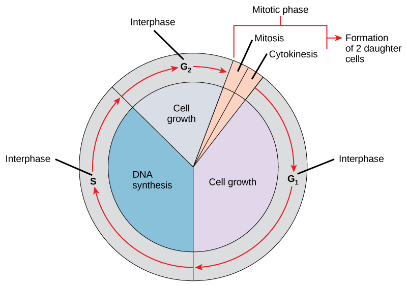
细胞周期是一系列有序事件,涉及细胞生长和细胞分裂,产生两个新的子细胞。 细胞分裂道路上的细胞经历了一系列精确定时且经过精心调节的生长、DNA复制和分裂阶段,这些阶段会产生两个相同的(克隆)细胞。 细胞周期有两个主要阶段:间期和有丝分裂期(图\(\PageIndex{1}\))。 在中间阶段,细胞生长,DNA被复制。 在有丝分裂阶段,复制的DNA和细胞质内容被分离,细胞分裂。
相间
在中间阶段,细胞经历正常的生长过程,同时也为细胞分裂做准备。 为了使细胞从中间阶段进入有丝分裂阶段,必须满足许多内部和外部条件。 相间的三个阶段称为 G 1、S 和 G 2。
G 1 阶段(第一间隙)
相间的第一阶段被称为 G 1 相(第一间隙),因为从微观角度来看,几乎看不到变化。 但是,在G 1 阶段,细胞在生化水平上非常活跃。 该细胞正在积累染色体 DNA 和相关蛋白质的组成部分,并积累足够的能量储备,以完成复制细胞核中每条染色体的任务。
S 相(DNA 的合成)
在整个中间阶段,核 DNA 保持半浓缩的染色质构型。 在 S 阶段,DNA 复制可以通过机制进行,这些机制会形成牢固地附着在质心区域上的相同的 DNA 分子对(姐妹染色体)。 中心体在 S 阶段被复制。 这两个中心体将产生有丝分裂主轴,这是在有丝分裂期间协调染色体运动的设备。 在每个动物细胞的中心,动物细胞的中心体与一对相互成直角的棒状物体,即中心体相关联。 中心体有助于组织细胞分裂。 中心体不存在于其他真核生物物种(例如植物和大多数真菌)的中心体中。
G 2 阶段(第二间隙)
在 G 2 阶段,细胞补充其能量储存并合成操纵染色体所需的蛋白质。 一些细胞器是复制的,细胞骨架被拆除,为有丝分裂阶段提供资源。 在 G 2 期间可能会有额外的细胞生长。 在细胞进入有丝分裂的第一阶段之前,必须完成有丝分裂阶段的最后准备。
有丝分裂阶段
有丝分裂阶段是一个多步骤过程,在此过程中,重复的染色体对齐、分离并移入两个新的相同子细胞。 有丝分裂阶段的第一部分称为核分裂或核分裂。 有丝分裂阶段的第二部分称为细胞分裂,是将细胞质成分物理分离成两个子细胞。
核分裂症(有丝分裂)
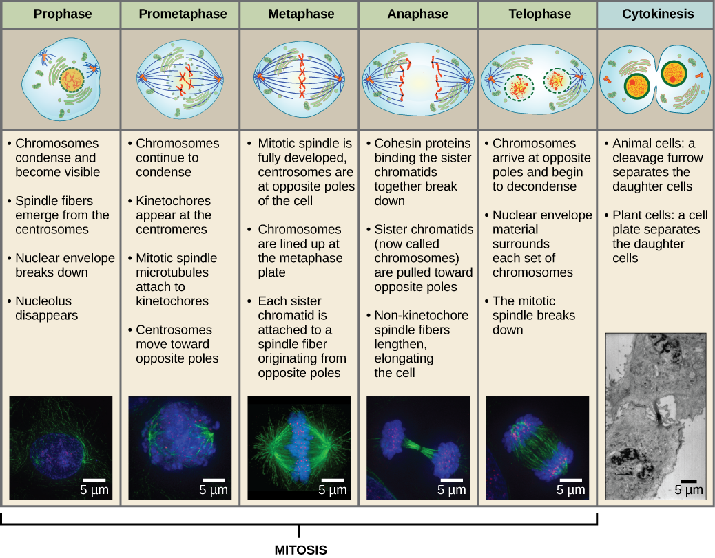
Karyokines is,也称为有丝分裂,分为一系列阶段(前期、前期、中期、后期和终期),这些阶段导致细胞核分裂(图\(\PageIndex{2}\)). Karyokinesis is also called mitosis.
Exercise \(\PageIndex{1}\)
Which of the following is the correct order of events in mitosis?
- Sister chromatids line up at the metaphase plate. The kinetochore becomes attached to the mitotic spindle. The nucleus reforms and the cell divides. Cohesin proteins break down and the sister chromatids separate.
- The kinetochore becomes attached to the mitotic spindle. Cohesin proteins break down and the sister chromatids separate. Sister chromatids line up at the metaphase plate. The nucleus reforms and the cell divides.
- The kinetochore becomes attached to the cohesin proteins. Sister chromatids line up at the metaphase plate. The kinetochore breaks down and the sister chromatids separate. The nucleus reforms and the cell divides.
- The kinetochore becomes attached to the mitotic spindle. Sister chromatids line up at the metaphase plate. Cohesin proteins break down and the sister chromatids separate. The nucleus reforms and the cell divides.
- Answer
-
D. The kinetochore becomes attached to the mitotic spindle. Sister chromatids line up at the metaphase plate. Cohesin proteins break down and the sister chromatids separate. The nucleus reforms and the cell divides.
During prophase, the “first phase,” the nuclear envelope starts to dissociate into small vesicles, and the membranous organelles (such as the Golgi complex or Golgi apparatus, and endoplasmic reticulum), fragment and disperse toward the periphery of the cell. The nucleolus disappears (disperses). The centrosomes begin to move to opposite poles of the cell. Microtubules that will form the mitotic spindle extend between the centrosomes, pushing them farther apart as the microtubule fibers lengthen. The sister chromatids begin to coil more tightly with the aid of condensin proteins and become visible under a light microscope.
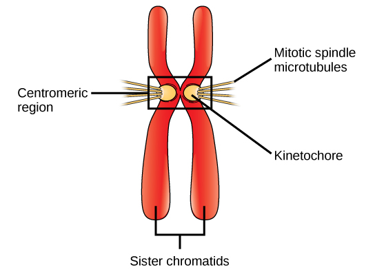
During prometaphase, the “first change phase,” many processes that were begun in prophase continue to advance. The remnants of the nuclear envelope fragment. The mitotic spindle continues to develop as more microtubules assemble and stretch across the length of the former nuclear area. Chromosomes become more condensed and discrete. Each sister chromatid develops a protein structure called a kinetochore in the centromeric region (Figure \(\PageIndex{3}\)). The proteins of the kinetochore attract and bind mitotic spindle microtubules. As the spindle microtubules extend from the centrosomes, some of these microtubules come into contact with and firmly bind to the kinetochores. Once a mitotic fiber attaches to a chromosome, the chromosome will be oriented until the kinetochores of sister chromatids face the opposite poles. Eventually, all the sister chromatids will be attached via their kinetochores to microtubules from opposing poles. Spindle microtubules that do not engage the chromosomes are called polar microtubules. These microtubules overlap each other midway between the two poles and contribute to cell elongation. Astral microtubules are located near the poles, aid in spindle orientation, and are required for the regulation of mitosis.
During metaphase, the “change phase,” all the chromosomes are aligned in a plane called the metaphase plate, or the equatorial plane, midway between the two poles of the cell. The sister chromatids are still tightly attached to each other by cohesin proteins. At this time, the chromosomes are maximally condensed.
During anaphase, the “upward phase,” the cohesin proteins degrade, and the sister chromatids separate at the centromere. Each chromatid, now called a chromosome, is pulled rapidly toward the centrosome to which its microtubule is attached. The cell becomes visibly elongated (oval shaped) as the polar microtubules slide against each other at the metaphase plate where they overlap.
During telophase, the “distance phase,” the chromosomes reach the opposite poles and begin to decondense (unravel), relaxing into a chromatin configuration. The mitotic spindles are depolymerized into tubulin monomers that will be used to assemble cytoskeletal components for each daughter cell. Nuclear envelopes form around the chromosomes, and nucleosomes appear within the nuclear area.
Cytokinesis
Cytokinesis, or “cell motion,” is the second main stage of the mitotic phase during which cell division is completed via the physical separation of the cytoplasmic components into two daughter cells. Division is not complete until the cell components have been apportioned and completely separated into the two daughter cells. Although the stages of mitosis are similar for most eukaryotes, the process of cytokinesis is quite different for eukaryotes that have cell walls, such as plant cells.
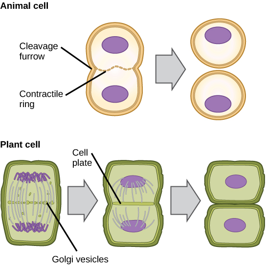
In cells such as animal cells that lack cell walls, cytokinesis follows the onset of anaphase. A contractile ring composed of actin filaments forms just inside the plasma membrane at the former metaphase plate. The actin filaments pull the equator of the cell inward, forming a fissure. This fissure, or “crack,” is called the cleavage furrow. The furrow deepens as the actin ring contracts, and eventually the membrane is cleaved in two (Figure \(\PageIndex{4}\)).
In plant cells, a new cell wall must form between the daughter cells. During interphase, the Golgi apparatus accumulates enzymes, structural proteins, and glucose molecules prior to breaking into vesicles and dispersing throughout the dividing cell. During telophase, these Golgi vesicles are transported on microtubules to form a phragmoplast (a vesicular structure) at the metaphase plate. There, the vesicles fuse and coalesce from the center toward the cell walls; this structure is called a cell plate. As more vesicles fuse, the cell plate enlarges until it merges with the cell walls at the periphery of the cell. Enzymes use the glucose that has accumulated between the membrane layers to build a new cell wall. The Golgi membranes become parts of the plasma membrane on either side of the new cell wall (Figure \(\PageIndex{4}\)).
G0 Phase
Not all cells adhere to the classic cell cycle pattern in which a newly formed daughter cell immediately enters the preparatory phases of interphase, closely followed by the mitotic phase. Cells in G0 phase are not actively preparing to divide. The cell is in a quiescent (inactive) stage that occurs when cells exit the cell cycle. Some cells enter G0 temporarily until an external signal triggers the onset of G1. Other cells that never or rarely divide, such as mature cardiac muscle and nerve cells, remain in G0 permanently.
Scientific Method Connection: Determine the Time Spent in Cell Cycle Stages
Problem: How long does a cell spend in interphase compared to each stage of mitosis?
Background: A prepared microscope slide of blastula cross-sections will show cells arrested in various stages of the cell cycle. It is not visually possible to separate the stages of interphase from each other, but the mitotic stages are readily identifiable. If 100 cells are examined, the number of cells in each identifiable cell cycle stage will give an estimate of the time it takes for the cell to complete that stage.
Problem Statement: Given the events included in all of interphase and those that take place in each stage of mitosis, estimate the length of each stage based on a 24-hour cell cycle. Before proceeding, state your hypothesis.
Test your hypothesis: Test your hypothesis by doing the following:
- Place a fixed and stained microscope slide of whitefish blastula cross-sections under the scanning objective of a light microscope.
- Locate and focus on one of the sections using the scanning objective of your microscope. Notice that the section is a circle composed of dozens of closely packed individual cells.
- Switch to the low-power objective and refocus. With this objective, individual cells are visible.
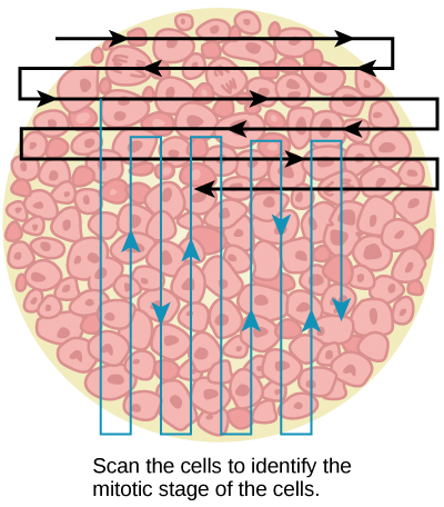
- Switch to the high-power objective and slowly move the slide left to right, and up and down to view all the cells in the section (Figure \(\PageIndex{5}\)). As you scan, you will notice that most of the cells are not undergoing mitosis but are in the interphase period of the cell cycle.
(a) 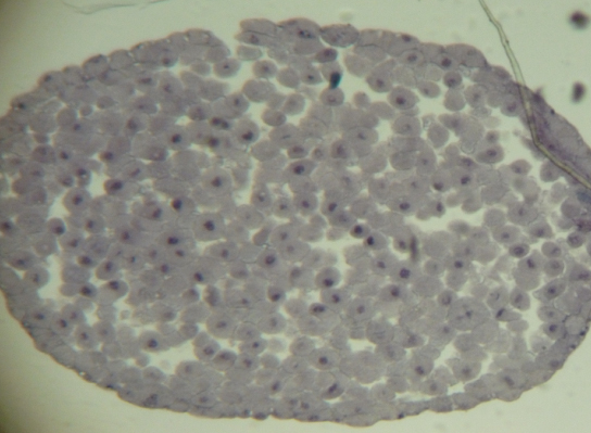
(b) Figure \(\PageIndex{5}\): Slowly scan whitefish blastula cells with the high-power objective as illustrated in image (a) to identify their mitotic stage. (b) A microscopic image of the scanned cells is shown. (credit “micrograph”: modification of work by Linda Flora; scale-bar data from Matt Russell) -
Practice identifying the various stages of the cell cycle, using the drawings of the stages as a guide (Figure \(\PageIndex{2}\)).
-
Once you are confident about your identification, begin to record the stage of each cell you encounter as you scan left to right, and top to bottom across the blastula section.
-
Keep a tally of your observations and stop when you reach 100 cells identified.
-
The larger the sample size (total number of cells counted), the more accurate the results. If possible, gather and record group data prior to calculating percentages and making estimates.
Record your observations: Make a table similar to the Table \(\PageIndex{1}\) in which you record your observations.
Table \(\PageIndex{1}\): Results of cell stage identification.
| Phase or Stage | Individual Totals | Group Totals | Percent |
|---|---|---|---|
| Interphase | |||
| Prophase | |||
| Metaphase | |||
| Anaphase | |||
| Telophase | |||
| Cytokinesis | |||
| Totals | 100 | 100 | 100 percent |
Analyze your data/report your results: To find the length of time whitefish blastula cells spend in each stage, multiply the percent (recorded as a decimal) by 24 hours. Make a table similar to the Table \(\PageIndex{2}\) to illustrate your data.
Table \(\PageIndex{2}\): Estimate of cell stage length.
| Phase or Stage | Percent (as Decimal) | Time in Hours |
|---|---|---|
| Interphase | ||
| Prophase | ||
| Metaphase | ||
| Anaphase | ||
| Telophase | ||
| Cytokinesis |
Draw a conclusion: Did your results support your estimated times? Were any of the outcomes unexpected? If so, discuss which events in that stage might contribute to the calculated time.
Summary
The cell cycle is an orderly sequence of events. Cells on the path to cell division proceed through a series of precisely timed and carefully regulated stages. In eukaryotes, the cell cycle consists of a long preparatory period, called interphase. Interphase is divided into G1, S, and G2 phases. The mitotic phase begins with karyokinesis (mitosis), which consists of five stages: prophase, prometaphase, metaphase, anaphase, and telophase. The final stage of the mitotic phase is cytokinesis, during which the cytoplasmic components of the daughter cells are separated either by an actin ring (animal cells) or by cell plate formation (plant cells).
Glossary
- anaphase
- stage of mitosis during which sister chromatids are separated from each other
- cell cycle
- ordered series of events involving cell growth and cell division that produces two new daughter cells
- cell plate
- structure formed during plant cell cytokinesis by Golgi vesicles, forming a temporary structure (phragmoplast) and fusing at the metaphase plate; ultimately leads to the formation of cell walls that separate the two daughter cells
- centriole
- rod-like structure constructed of microtubules at the center of each animal cell centrosome
- cleavage furrow
- constriction formed by an actin ring during cytokinesis in animal cells that leads to cytoplasmic division
- condensin
- proteins that help sister chromatids coil during prophase
- cytokinesis
- division of the cytoplasm following mitosis that forms two daughter cells.
- G0 phase
- distinct from the G1 phase of interphase; a cell in G0 is not preparing to divide
- G1 phase
- (also, first gap) first phase of interphase centered on cell growth during mitosis
- G2 phase
- (also, second gap) third phase of interphase during which the cell undergoes final preparations for mitosis
- interphase
- period of the cell cycle leading up to mitosis; includes G1, S, and G2 phases (the interim period between two consecutive cell divisions
- karyokinesis
- mitotic nuclear division
- kinetochore
- protein structure associated with the centromere of each sister chromatid that attracts and binds spindle microtubules during prometaphase
- metaphase plate
- equatorial plane midway between the two poles of a cell where the chromosomes align during metaphase
- metaphase
- stage of mitosis during which chromosomes are aligned at the metaphase plate
- mitosis
- (also, karyokinesis) period of the cell cycle during which the duplicated chromosomes are separated into identical nuclei; includes prophase, prometaphase, metaphase, anaphase, and telophase
- mitotic phase
- period of the cell cycle during which duplicated chromosomes are distributed into two nuclei and cytoplasmic contents are divided; includes karyokinesis (mitosis) and cytokinesis
- mitotic spindle
- apparatus composed of microtubules that orchestrates the movement of chromosomes during mitosis
- prometaphase
- stage of mitosis during which the nuclear membrane breaks down and mitotic spindle fibers attach to kinetochores
- prophase
- stage of mitosis during which chromosomes condense and the mitotic spindle begins to form
- quiescent
- refers to a cell that is performing normal cell functions and has not initiated preparations for cell division
- S phase
- second, or synthesis, stage of interphase during which DNA replication occurs
- telophase
- stage of mitosis during which chromosomes arrive at opposite poles, decondense, and are surrounded by a new nuclear envelope


