
35.3: מערכת העצבים המרכזית
- Page ID
- 205935

מיומנויות לפיתוח
- זהה את חוט השדרה, אונות המוח ואזורי מוח אחרים בתרשים של המוח
- תאר את הפונקציות הבסיסיות של חוט השדרה, אונות המוח ואזורי מוח אחרים
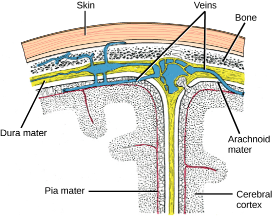
מערכת העצבים המרכזית (CNS) מורכבת מהמוח, שחלק ממנו מוצג באיור \(\PageIndex{1}\) ובחוט השדרה ומכוסה בשלוש שכבות של כיסויי מגן הנקראים קרום המוח (מהמילה היוונית לממברנה). השכבה החיצונית ביותר היא הדורה מאטר (בלטינית "אם קשה"). כפי שמציע הלטינית, התפקיד העיקרי לשכבה עבה זו הוא להגן על המוח ועל חוט השדרה. הדורה מאטר מכילה גם מבנים דמויי ורידים המובילים דם מהמוח חזרה ללב. השכבה האמצעית היא החומר הארכנואיד דמוי הרשת. השכבה האחרונה היא pia mater (בלטינית עבור "אמא רכה"), אשר יוצר קשר ישיר ומכסה את המוח וחוט השדרה כמו ניילון נצמד. החלל בין הארכנואיד לפיא מאטר מלא בנוזל מוחי (CSF). CSF מיוצר על ידי רקמה הנקראת מקלעת כורואיד בתאים מלאי נוזלים במערכת העצבים המרכזית הנקראת חדרים. המוח צף ב- CSF, הפועל ככרית ובולם זעזועים והופך את המוח לצוף ניטרלי. CSF מתפקד גם להפצת חומרים כימיים ברחבי המוח ולחוט השדרה.
המוח כולו מכיל רק כ 8.5 כפות CSF, אך CSF מיוצר כל הזמן בחדרים. זה יוצר בעיה כאשר חדר חסום - ה- CSF מצטבר ויוצר נפיחות והמוח נדחף אל הגולגולת. מצב נפיחות זה נקרא הידרוצפלוס ("ראש מים") ויכול לגרום להתקפים, בעיות קוגניטיביות ואפילו מוות אם לא מוחדר שאנט להסרת הנוזל והלחץ.
המוח
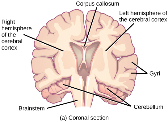
המוח הוא החלק של מערכת העצבים המרכזית הכלול בחלל הגולגולת של הגולגולת. הוא כולל את קליפת המוח, המערכת הלימבית, הגרעינים הבסיסיים, התלמוס, ההיפותלמוס והמוח הקטן. ישנן שלוש דרכים שונות בהן ניתן לחלק מוח על מנת לצפות במבנים פנימיים: קטע sagittal חותך את המוח משמאל לימין, כפי שמוצג באיור \(\PageIndex{2}\) ב ', קטע קורונלי חותך את המוח מלפנים לאחור, כפי שמוצג באיור \(\PageIndex{2}\) א, וחתך אופקי חותך את המוח מלמעלה למטה.
קליפת המוח
החלק החיצוני ביותר של המוח הוא חתיכה עבה של רקמת מערכת העצבים הנקראת קליפת המוח, המקופלת לגבעות הנקראות gyri (יחיד: gyrus) ועמקים הנקראים sulci (יחיד: sulcus). קליפת המוח מורכבת משתי חצאי כדור - ימין ושמאל - המופרדות על ידי סולקוס גדול. צרור סיבים עבה הנקרא corpus callosum (בלטינית: "גוף קשוח") מחבר בין שתי ההמיספרות ומאפשר העברת מידע מצד אחד לצד השני. למרות שיש כמה תפקודי מוח הממוקמים יותר לחצי הכדור האחד מהשני, הפונקציות של שתי ההמיספרות מיותרות במידה רבה. למעשה, לפעמים (לעיתים רחוקות מאוד) מוסרים חצי כדור שלם לטיפול באפילפסיה קשה. בעוד שחולים אכן סובלים מליקויים מסוימים בעקבות הניתוח, הם עלולים להיתקל בבעיות מעטות באופן מפתיע, במיוחד כאשר הניתוח מבוצע בילדים שיש להם מערכת עצבים מאוד לא בשלה.
בניתוחים אחרים לטיפול באפילפסיה קשה, קורפוס הקאלוסום נחתך במקום להסיר חצי כדור שלם. זה גורם למצב שנקרא מוח מפוצל, המעניק תובנות לגבי תפקודים ייחודיים של שתי ההמיספרות. לדוגמה, כאשר אובייקט מוצג לשדה הראייה השמאלי של המטופלים, ייתכן שהם לא יוכלו לתת שם מילולי לאובייקט (ועשויים לטעון שלא ראו אובייקט כלל). הסיבה לכך היא שהקלט החזותי משדה הראייה השמאלי חוצה ונכנס לחצי הכדור הימני ואז אינו יכול לאותת למרכז הדיבור, שנמצא בדרך כלל בצד שמאל של המוח. למרבה הפלא, אם מטופל מפוצל מוח יתבקש להרים אובייקט ספציפי מתוך קבוצת אובייקטים ביד שמאל, המטופל יוכל לעשות זאת אך עדיין לא יוכל לזהות אותו קולית.
קישור ללמידה

עיין באתר זה כדי ללמוד עוד על חולי מוח מפוצל ולשחק משחק שבו תוכל לדגמן את הניסויים במוח המפוצל בעצמך.
כל חצי כדור קליפת המוח מכיל אזורים הנקראים אונות המעורבים בתפקודים שונים. מדענים משתמשים בטכניקות שונות כדי לקבוע אילו אזורי מוח מעורבים בתפקודים שונים: הם בוחנים חולים שסבלו מפציעות או מחלות המשפיעות על אזורים ספציפיים ורואים כיצד אזורים אלה קשורים לחסרים תפקודיים. הם גם עורכים מחקרים בבעלי חיים בהם הם מעוררים אזורי מוח ורואים אם יש שינויים התנהגותיים. הם משתמשים בטכניקה הנקראת גירוי טרנסמגנטי (TMS) כדי להשבית באופן זמני חלקים ספציפיים בקליפת המוח באמצעות מגנטים חזקים המונחים מחוץ לראש; והם משתמשים בהדמיית תהודה מגנטית פונקציונלית (fMRI) כדי לבחון שינויים בזרימת הדם המחומצנת באזורי מוח מסוימים המתואמים למשימות התנהגותיות ספציפיות. טכניקות אלה ואחרות העניקו תובנה רבה לתפקודים של אזורי מוח שונים, אך הראו גם כי כל אזור מוח נתון יכול להיות מעורב ביותר מהתנהגות או תהליך אחד, וכל התנהגות או תהליך נתון כרוך בדרך כלל נוירונים באזורים מוחיים מרובים.. עם זאת, ניתן לפרק כל חצי כדור של קליפת המוח של היונקים לארבע אונות מוגדרות תפקודית ומרחבית: חזיתית, פריאטלית, טמפורלית ואוקסיפיטלית. איור \(\PageIndex{3}\) illustrates these four lobes of the human cerebral cortex.
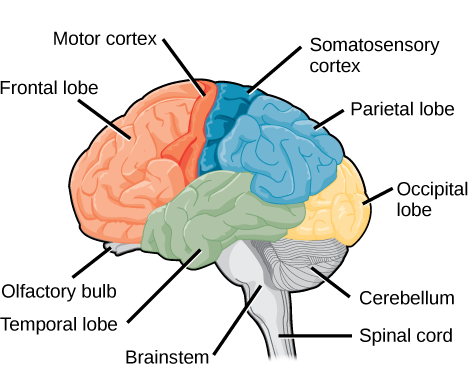
The frontal lobe is located at the front of the brain, over the eyes. This lobe contains the olfactory bulb, which processes smells. The frontal lobe also contains the motor cortex, which is important for planning and implementing movement. Areas within the motor cortex map to different muscle groups, and there is some organization to this map, as shown in Figure \(\PageIndex{4}\). For example, the neurons that control movement of the fingers are next to the neurons that control movement of the hand. Neurons in the frontal lobe also control cognitive functions like maintaining attention, speech, and decision-making. Studies of humans who have damaged their frontal lobes show that parts of this area are involved in personality, socialization, and assessing risk.
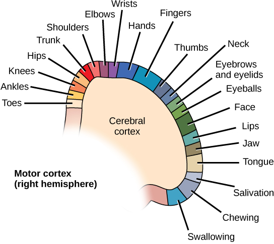
The parietal lobe is located at the top of the brain. Neurons in the parietal lobe are involved in speech and also reading. Two of the parietal lobe’s main functions are processing somatosensation—touch sensations like pressure, pain, heat, cold—and processing proprioception—the sense of how parts of the body are oriented in space. The parietal lobe contains a somatosensory map of the body similar to the motor cortex.
The occipital lobe is located at the back of the brain. It is primarily involved in vision—seeing, recognizing, and identifying the visual world.
The temporal lobe is located at the base of the brain by your ears and is primarily involved in processing and interpreting sounds. It also contains the hippocampus (Greek for “seahorse”)—a structure that processes memory formation. The hippocampus is illustrated in Figure \(\PageIndex{6}\). The role of the hippocampus in memory was partially determined by studying one famous epileptic patient, HM, who had both sides of his hippocampus removed in an attempt to cure his epilepsy. His seizures went away, but he could no longer form new memories (although he could remember some facts from before his surgery and could learn new motor tasks).
Evolution Connection: Cerebral Cortex
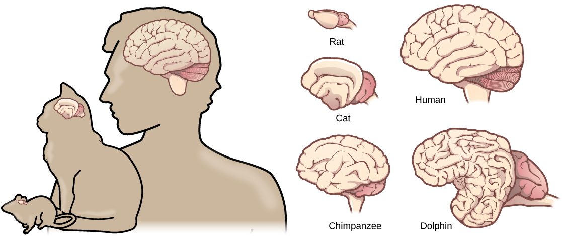
Compared to other vertebrates, mammals have exceptionally large brains for their body size. An entire alligator’s brain, for example, would fill about one and a half teaspoons. This increase in brain to body size ratio is especially pronounced in apes, whales, and dolphins. While this increase in overall brain size doubtlessly played a role in the evolution of complex behaviors unique to mammals, it does not tell the whole story. Scientists have found a relationship between the relatively high surface area of the cortex and the intelligence and complex social behaviors exhibited by some mammals. This increased surface area is due, in part, to increased folding of the cortical sheet (more sulci and gyri). For example, a rat cortex is very smooth with very few sulci and gyri. Cat and sheep cortices have more sulci and gyri. Chimps, humans, and dolphins have even more.
Basal Ganglia
Interconnected brain areas called the basal ganglia (or basal nuclei) play important roles in movement control and posture. Damage to the basal ganglia, as in Parkinson’s disease, leads to motor impairments like a shuffling gait when walking. The basal ganglia also regulate motivation. For example, when a wasp sting led to bilateral basal ganglia damage in a 25-year-old businessman, he began to spend all his days in bed and showed no interest in anything or anybody. But when he was externally stimulated—as when someone asked to play a card game with him—he was able to function normally. Interestingly, he and other similar patients do not report feeling bored or frustrated by their state.
Thalamus
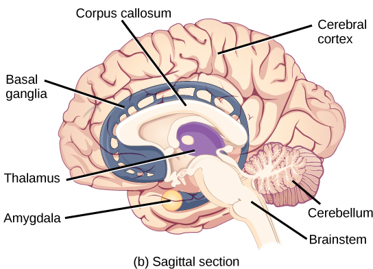
The thalamus (Greek for “inner chamber”), illustrated in Figure \(\PageIndex{6}\), acts as a gateway to and from the cortex. It receives sensory and motor inputs from the body and also receives feedback from the cortex. This feedback mechanism can modulate conscious awareness of sensory and motor inputs depending on the attention and arousal state of the animal. The thalamus helps regulate consciousness, arousal, and sleep states. A rare genetic disorder called fatal familial insomnia causes the degeneration of thalamic neurons and glia. This disorder prevents affected patients from being able to sleep, among other symptoms, and is eventually fatal.

Hypothalamus
Below the thalamus is the hypothalamus, shown in Figure \(\PageIndex{6}\). The hypothalamus controls the endocrine system by sending signals to the pituitary gland, a pea-sized endocrine gland that releases several different hormones that affect other glands as well as other cells. This relationship means that the hypothalamus regulates important behaviors that are controlled by these hormones. The hypothalamus is the body’s thermostat—it makes sure key functions like food and water intake, energy expenditure, and body temperature are kept at appropriate levels. Neurons within the hypothalamus also regulate circadian rhythms, sometimes called sleep cycles.
Limbic System
The limbic system is a connected set of structures that regulates emotion, as well as behaviors related to fear and motivation. It plays a role in memory formation and includes parts of the thalamus and hypothalamus as well as the hippocampus. One important structure within the limbic system is a temporal lobe structure called the amygdala (Greek for “almond”), illustrated in Figure \(\PageIndex{6}\). The two amygdala are important both for the sensation of fear and for recognizing fearful faces. The cingulate gyrus helps regulate emotions and pain.
Cerebellum
The cerebellum (Latin for “little brain”), shown in Figure \(\PageIndex{3}\), sits at the base of the brain on top of the brainstem. The cerebellum controls balance and aids in coordinating movement and learning new motor tasks.
Brainstem
The brainstem, illustrated in Figure \(\PageIndex{3}\), connects the rest of the brain with the spinal cord. It consists of the midbrain, medulla oblongata, and the pons. Motor and sensory neurons extend through the brainstem allowing for the relay of signals between the brain and spinal cord. Ascending neural pathways cross in this section of the brain allowing the left hemisphere of the cerebrum to control the right side of the body and vice versa. The brainstem coordinates motor control signals sent from the brain to the body. The brainstem controls several important functions of the body including alertness, arousal, breathing, blood pressure, digestion, heart rate, swallowing, walking, and sensory and motor information integration.
Spinal Cord
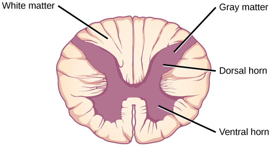
Connecting to the brainstem and extending down the body through the spinal column is the spinal cord, shown in Figure \(\PageIndex{3}\). The spinal cord is a thick bundle of nerve tissue that carries information about the body to the brain and from the brain to the body. The spinal cord is contained within the bones of the vertebrate column but is able to communicate signals to and from the body through its connections with spinal nerves (part of the peripheral nervous system). A cross-section of the spinal cord looks like a white oval containing a gray butterfly-shape, as illustrated in Figure \(\PageIndex{7}\). Myelinated axons make up the “white matter” and neuron and glial cell bodies make up the “gray matter.” Gray matter is also composed of interneurons, which connect two neurons each located in different parts of the body. Axons and cell bodies in the dorsal (facing the back of the animal) spinal cord convey mostly sensory information from the body to the brain. Axons and cell bodies in the ventral (facing the front of the animal) spinal cord primarily transmit signals controlling movement from the brain to the body.
The spinal cord also controls motor reflexes. These reflexes are quick, unconscious movements—like automatically removing a hand from a hot object. Reflexes are so fast because they involve local synaptic connections. For example, the knee reflex that a doctor tests during a routine physical is controlled by a single synapse between a sensory neuron and a motor neuron. While a reflex may only require the involvement of one or two synapses, synapses with interneurons in the spinal column transmit information to the brain to convey what happened (the knee jerked, or the hand was hot).
In the United States, there around 10,000 spinal cord injuries each year. Because the spinal cord is the information superhighway connecting the brain with the body, damage to the spinal cord can lead to paralysis. The extent of the paralysis depends on the location of the injury along the spinal cord and whether the spinal cord was completely severed. For example, if the spinal cord is damaged at the level of the neck, it can cause paralysis from the neck down, whereas damage to the spinal column further down may limit paralysis to the legs. Spinal cord injuries are notoriously difficult to treat because spinal nerves do not regenerate, although ongoing research suggests that stem cell transplants may be able to act as a bridge to reconnect severed nerves. Researchers are also looking at ways to prevent the inflammation that worsens nerve damage after injury. One such treatment is to pump the body with cold saline to induce hypothermia. This cooling can prevent swelling and other processes that are thought to worsen spinal cord injuries.
Summary
The vertebrate central nervous system contains the brain and the spinal cord, which are covered and protected by three meninges. The brain contains structurally and functionally defined regions. In mammals, these include the cortex (which can be broken down into four primary functional lobes: frontal, temporal, occipital, and parietal), basal ganglia, thalamus, hypothalamus, limbic system, cerebellum, and brainstem—although structures in some of these designations overlap. While functions may be primarily localized to one structure in the brain, most complex functions, like language and sleep, involve neurons in multiple brain regions. The spinal cord is the information superhighway that connects the brain with the rest of the body through its connections with peripheral nerves. It transmits sensory and motor input and also controls motor reflexes.
Glossary
- amygdala
- structure within the limbic system that processes fear
- arachnoid mater
- spiderweb-like middle layer of the meninges that cover the central nervous system
- basal ganglia
- interconnected collections of cells in the brain that are involved in movement and motivation; also known as basal nuclei
- basal nuclei
- see basal ganglia
- brainstem
- portion of the brain that connects with the spinal cord; controls basic nervous system functions like breathing, heart rate, and swallowing
- cerebellum
- brain structure involved in posture, motor coordination, and learning new motor actions
- cerebral cortex
- outermost sheet of brain tissue; involved in many higher-order functions
- choroid plexus
- spongy tissue within ventricles that produces cerebrospinal fluid
- cingulate gyrus
- helps regulate emotions and pain; thought to directly drive the body’s conscious response to unpleasant experiences
- corpus callosum
- thick fiber bundle that connects the cerebral hemispheres
- cerebrospinal fluid (CSF)
- clear liquid that surrounds the brain and spinal cord and fills the ventricles and central canal; acts as a shock absorber and circulates material throughout the brain and spinal cord.
- dura mater
- tough outermost layer that covers the central nervous system
- frontal lobe
- part of the cerebral cortex that contains the motor cortex and areas involved in planning, attention, and language
- gyrus
- (plural: gyri) ridged protrusions in the cortex
- hippocampus
- brain structure in the temporal lobe involved in processing memories
- hypothalamus
- brain structure that controls hormone release and body homeostasis
- limbic system
- connected brain areas that process emotion and motivation
- meninge
- membrane that covers and protects the central nervous system
- occipital lobe
- part of the cerebral cortex that contains visual cortex and processes visual stimuli
- parietal lobe
- part of the cerebral cortex involved in processing touch and the sense of the body in space
- pia mater
- thin membrane layer directly covering the brain and spinal cord
- proprioception
- sense about how parts of the body are oriented in space
- somatosensation
- sense of touch
- spinal cord
- thick fiber bundle that connects the brain with peripheral nerves; transmits sensory and motor information; contains neurons that control motor reflexes
- sulcus
- (plural: sulci) indents or “valleys” in the cortex
- temporal lobe
- part of the cerebral cortex that processes auditory input; parts of the temporal lobe are involved in speech, memory, and emotion processing
- thalamus
- brain area that relays sensory information to the cortex
- ventricle
- cavity within brain that contains cerebrospinal fluid