
35.3: Mfumo wa neva wa Kati
- Page ID
- 176031

Ujuzi wa Kuendeleza
- Tambua kamba ya mgongo, lobes ya ubongo, na maeneo mengine ya ubongo kwenye mchoro wa ubongo
- Eleza kazi za msingi za kamba ya mgongo, lobes ya ubongo, na maeneo mengine ya ubongo
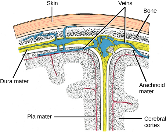
Mfumo mkuu wa neva (CNS) hujumuisha ubongo, sehemu ambayo inavyoonekana kwenye Kielelezo\(\PageIndex{1}\) na kamba ya mgongo na inafunikwa na tabaka tatu za vifuniko vya kinga vinavyoitwa meninges (kutoka neno la Kigiriki kwa membrane). Safu ya nje ni mama wa kudumu (Kilatini kwa “mama mgumu”). Kama Kilatini inavyoonyesha, kazi ya msingi kwa safu hii nyembamba ni kulinda ubongo na kamba ya mgongo. Mater ya kudumu pia ina miundo kama mshipa ambayo hubeba damu kutoka ubongo kurudi moyoni. Safu ya kati ni mtandao-kama arachnoid mater. Safu ya mwisho ni pia mater (Kilatini kwa “mama laini”), ambayo huwasiliana moja kwa moja na inashughulikia ubongo na uti wa mgongo kama ukingo wa plastiki. Nafasi kati ya maters ya arachnoid na pia imejaa maji ya cerebrospinal (CSF). CSF huzalishwa na tishu inayoitwa plexus ya choroid katika vyumba vilivyojaa maji katika CNS inayoitwa ventricles. Ubongo unaelea katika CSF, ambayo hufanya kama mto na mshtuko wa mshtuko na hufanya ubongo usiwe na buoyant. CSF pia inafanya kazi ya kuzunguka vitu vya kemikali katika ubongo na ndani ya kamba ya mgongo.
Ubongo mzima una vijiko 8.5 tu vya CSF, lakini CSF inazalishwa mara kwa mara katika ventricles. Hii inajenga tatizo wakati ventricle imezuwa-CSF hujenga na inajenga uvimbe na ubongo unasukumwa dhidi ya fuvu. Hali hii ya uvimbe inaitwa hydrocephalus (“kichwa cha maji”) na inaweza kusababisha mshtuko, matatizo ya utambuzi, na hata kifo ikiwa shunt haiingiziwi ili kuondoa maji na shinikizo.
Ubongo
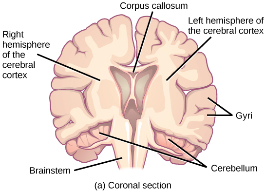
Ubongo ni sehemu ya mfumo mkuu wa neva unao kwenye cavity ya fuvu ya fuvu. Inajumuisha kamba ya ubongo, mfumo wa limbic, ganglia ya basal, thalamus, hypothalamus, na cerebellum. Kuna njia tatu tofauti ambazo ubongo unaweza kugawanywa ili kuona miundo ya ndani: sehemu ya sagittal kupunguzwa ubongo kushoto kwenda kulia, kama inavyoonekana katika Kielelezo\(\PageIndex{2}\) b, sehemu ya coronal hupunguza ubongo mbele kwa nyuma, kama inavyoonekana katika Kielelezo\(\PageIndex{2}\) a, na sehemu ya usawa kupunguzwa ubongo juu hadi chini.
Kamba ya ubongo
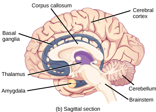
Sehemu ya nje ya ubongo ni kipande kikubwa cha tishu za mfumo wa neva kinachoitwa gamba la ubongo, ambalo linaingizwa kwenye milima inayoitwa gyri (umoja: gyrus) na mabonde yanayoitwa sulci (umoja: sulcus). Gamba linaundwa na hemispheres mbili—kulia na kushoto—ambazo zinajitenga na sulcus kubwa. Kifungu kikubwa cha nyuzi kinachoitwa corpus callosum (Kilatini: “mwili mgumu”) huunganisha hemispheres mbili na inaruhusu habari kupitishwa kutoka upande mmoja hadi mwingine. Ingawa kuna baadhi ya kazi za ubongo ambazo zinakaa zaidi kwa hemisphere moja kuliko nyingine, kazi za hemispheres mbili ni nyingi sana. Kwa kweli, wakati mwingine (mara chache sana) hemisphere nzima imeondolewa kutibu kifafa kali. Wakati wagonjwa wanakabiliwa na upungufu kufuatia upasuaji, wanaweza kuwa na matatizo ya kushangaza machache, hasa wakati upasuaji unafanywa kwa watoto ambao wana mifumo ya neva machanga sana.
Katika upasuaji mwingine kutibu kifafa kali, callosum corpus hukatwa badala ya kuondoa hemisphere nzima. Hii inasababisha hali inayoitwa split-ubongo, ambayo inatoa ufahamu katika kazi za kipekee za hemispheres mbili. Kwa mfano, wakati kitu kinapowasilishwa kwa uwanja wa kuona wa kushoto wa wagonjwa, huenda hawawezi kutaja kitu kwa maneno (na wanaweza kudai kuwa hawajaona kitu kabisa). Hii ni kwa sababu pembejeo ya kuona kutoka shamba la kuona la kushoto linavuka na kuingia katika ulimwengu wa kulia na hauwezi kuashiria kituo cha hotuba, ambacho kwa ujumla hupatikana upande wa kushoto wa ubongo. Kwa kushangaza, ikiwa mgonjwa wa mgawanyiko wa ubongo anaulizwa kuchukua kitu maalum nje ya kikundi cha vitu na mkono wa kushoto, mgonjwa ataweza kufanya hivyo lakini bado hawezi kuitambua kwa sauti.
Unganisha na Kujifunza

Angalia tovuti hii ili ujifunze zaidi kuhusu wagonjwa wa mgawanyiko wa ubongo na kucheza mchezo ambapo unaweza kuiga majaribio ya ubongo wa mgawanyiko mwenyewe.
Kila hekta ya kamba ina mikoa inayoitwa lobes ambayo inahusika katika kazi tofauti. Wanasayansi hutumia mbinu mbalimbali kuamua maeneo gani ya ubongo yanahusika katika kazi tofauti: huchunguza wagonjwa ambao wamekuwa na majeraha au magonjwa yanayoathiri maeneo maalum na kuona jinsi maeneo hayo yanahusiana na upungufu wa kazi. Pia hufanya masomo ya wanyama ambapo huchochea maeneo ya ubongo na kuona kama kuna mabadiliko yoyote ya tabia. Wanatumia mbinu inayoitwa kusisimua transmagnetic (TMS) ili kuzima muda sehemu maalum za gamba kwa kutumia sumaku kali zilizowekwa nje ya kichwa; na wanatumia upigaji picha wa resonance magnetic kazi (fMRI) kuangalia mabadiliko katika mtiririko wa damu oksijeni katika maeneo fulani ya ubongo yanayohusiana na kazi maalum ya tabia. Mbinu hizi, na zingine, zimetoa ufahamu mkubwa katika kazi za mikoa mbalimbali ya ubongo lakini pia zimeonyesha kuwa eneo lolote la ubongo linaweza kuhusishwa katika tabia au mchakato zaidi ya moja, na tabia au mchakato wowote unaotolewa kwa ujumla unahusisha neuroni katika maeneo mbalimbali ya ubongo. Hiyo inasemekana, kila ulimwengu wa kamba ya ubongo wa mamalia inaweza kuvunjwa ndani ya lobes nne za kazi na za anga: mbele, parietali, temporal, na occipital. Kielelezo\(\PageIndex{3}\) illustrates these four lobes of the human cerebral cortex.
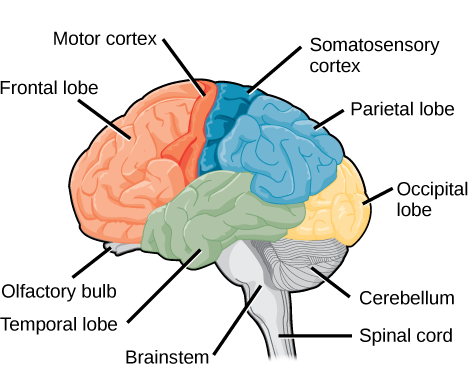
The frontal lobe is located at the front of the brain, over the eyes. This lobe contains the olfactory bulb, which processes smells. The frontal lobe also contains the motor cortex, which is important for planning and implementing movement. Areas within the motor cortex map to different muscle groups, and there is some organization to this map, as shown in Figure \(\PageIndex{4}\). For example, the neurons that control movement of the fingers are next to the neurons that control movement of the hand. Neurons in the frontal lobe also control cognitive functions like maintaining attention, speech, and decision-making. Studies of humans who have damaged their frontal lobes show that parts of this area are involved in personality, socialization, and assessing risk.
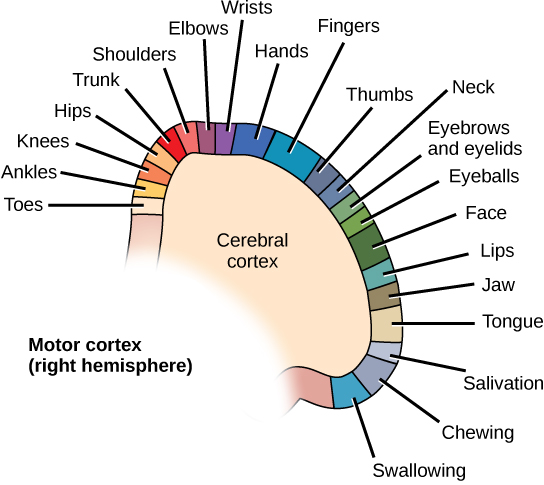
The parietal lobe is located at the top of the brain. Neurons in the parietal lobe are involved in speech and also reading. Two of the parietal lobe’s main functions are processing somatosensation—touch sensations like pressure, pain, heat, cold—and processing proprioception—the sense of how parts of the body are oriented in space. The parietal lobe contains a somatosensory map of the body similar to the motor cortex.
The occipital lobe is located at the back of the brain. It is primarily involved in vision—seeing, recognizing, and identifying the visual world.
The temporal lobe is located at the base of the brain by your ears and is primarily involved in processing and interpreting sounds. It also contains the hippocampus (Greek for “seahorse”)—a structure that processes memory formation. The hippocampus is illustrated in Figure \(\PageIndex{6}\). The role of the hippocampus in memory was partially determined by studying one famous epileptic patient, HM, who had both sides of his hippocampus removed in an attempt to cure his epilepsy. His seizures went away, but he could no longer form new memories (although he could remember some facts from before his surgery and could learn new motor tasks).
Evolution Connection: Cerebral Cortex
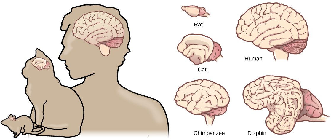
Compared to other vertebrates, mammals have exceptionally large brains for their body size. An entire alligator’s brain, for example, would fill about one and a half teaspoons. This increase in brain to body size ratio is especially pronounced in apes, whales, and dolphins. While this increase in overall brain size doubtlessly played a role in the evolution of complex behaviors unique to mammals, it does not tell the whole story. Scientists have found a relationship between the relatively high surface area of the cortex and the intelligence and complex social behaviors exhibited by some mammals. This increased surface area is due, in part, to increased folding of the cortical sheet (more sulci and gyri). For example, a rat cortex is very smooth with very few sulci and gyri. Cat and sheep cortices have more sulci and gyri. Chimps, humans, and dolphins have even more.
Basal Ganglia
Interconnected brain areas called the basal ganglia (or basal nuclei) play important roles in movement control and posture. Damage to the basal ganglia, as in Parkinson’s disease, leads to motor impairments like a shuffling gait when walking. The basal ganglia also regulate motivation. For example, when a wasp sting led to bilateral basal ganglia damage in a 25-year-old businessman, he began to spend all his days in bed and showed no interest in anything or anybody. But when he was externally stimulated—as when someone asked to play a card game with him—he was able to function normally. Interestingly, he and other similar patients do not report feeling bored or frustrated by their state.
Thalamus
The thalamus (Greek for “inner chamber”), illustrated in Figure \(\PageIndex{6}\), acts as a gateway to and from the cortex. It receives sensory and motor inputs from the body and also receives feedback from the cortex. This feedback mechanism can modulate conscious awareness of sensory and motor inputs depending on the attention and arousal state of the animal. The thalamus helps regulate consciousness, arousal, and sleep states. A rare genetic disorder called fatal familial insomnia causes the degeneration of thalamic neurons and glia. This disorder prevents affected patients from being able to sleep, among other symptoms, and is eventually fatal.

Hypothalamus
Below the thalamus is the hypothalamus, shown in Figure \(\PageIndex{6}\). The hypothalamus controls the endocrine system by sending signals to the pituitary gland, a pea-sized endocrine gland that releases several different hormones that affect other glands as well as other cells. This relationship means that the hypothalamus regulates important behaviors that are controlled by these hormones. The hypothalamus is the body’s thermostat—it makes sure key functions like food and water intake, energy expenditure, and body temperature are kept at appropriate levels. Neurons within the hypothalamus also regulate circadian rhythms, sometimes called sleep cycles.
Limbic System
The limbic system is a connected set of structures that regulates emotion, as well as behaviors related to fear and motivation. It plays a role in memory formation and includes parts of the thalamus and hypothalamus as well as the hippocampus. One important structure within the limbic system is a temporal lobe structure called the amygdala (Greek for “almond”), illustrated in Figure \(\PageIndex{6}\). The two amygdala are important both for the sensation of fear and for recognizing fearful faces. The cingulate gyrus helps regulate emotions and pain.
Cerebellum
The cerebellum (Latin for “little brain”), shown in Figure \(\PageIndex{3}\), sits at the base of the brain on top of the brainstem. The cerebellum controls balance and aids in coordinating movement and learning new motor tasks.
Brainstem
The brainstem, illustrated in Figure \(\PageIndex{3}\), connects the rest of the brain with the spinal cord. It consists of the midbrain, medulla oblongata, and the pons. Motor and sensory neurons extend through the brainstem allowing for the relay of signals between the brain and spinal cord. Ascending neural pathways cross in this section of the brain allowing the left hemisphere of the cerebrum to control the right side of the body and vice versa. The brainstem coordinates motor control signals sent from the brain to the body. The brainstem controls several important functions of the body including alertness, arousal, breathing, blood pressure, digestion, heart rate, swallowing, walking, and sensory and motor information integration.
Spinal Cord
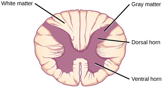
Connecting to the brainstem and extending down the body through the spinal column is the spinal cord, shown in Figure \(\PageIndex{3}\). The spinal cord is a thick bundle of nerve tissue that carries information about the body to the brain and from the brain to the body. The spinal cord is contained within the bones of the vertebrate column but is able to communicate signals to and from the body through its connections with spinal nerves (part of the peripheral nervous system). A cross-section of the spinal cord looks like a white oval containing a gray butterfly-shape, as illustrated in Figure \(\PageIndex{7}\). Myelinated axons make up the “white matter” and neuron and glial cell bodies make up the “gray matter.” Gray matter is also composed of interneurons, which connect two neurons each located in different parts of the body. Axons and cell bodies in the dorsal (facing the back of the animal) spinal cord convey mostly sensory information from the body to the brain. Axons and cell bodies in the ventral (facing the front of the animal) spinal cord primarily transmit signals controlling movement from the brain to the body.
The spinal cord also controls motor reflexes. These reflexes are quick, unconscious movements—like automatically removing a hand from a hot object. Reflexes are so fast because they involve local synaptic connections. For example, the knee reflex that a doctor tests during a routine physical is controlled by a single synapse between a sensory neuron and a motor neuron. While a reflex may only require the involvement of one or two synapses, synapses with interneurons in the spinal column transmit information to the brain to convey what happened (the knee jerked, or the hand was hot).
In the United States, there around 10,000 spinal cord injuries each year. Because the spinal cord is the information superhighway connecting the brain with the body, damage to the spinal cord can lead to paralysis. The extent of the paralysis depends on the location of the injury along the spinal cord and whether the spinal cord was completely severed. For example, if the spinal cord is damaged at the level of the neck, it can cause paralysis from the neck down, whereas damage to the spinal column further down may limit paralysis to the legs. Spinal cord injuries are notoriously difficult to treat because spinal nerves do not regenerate, although ongoing research suggests that stem cell transplants may be able to act as a bridge to reconnect severed nerves. Researchers are also looking at ways to prevent the inflammation that worsens nerve damage after injury. One such treatment is to pump the body with cold saline to induce hypothermia. This cooling can prevent swelling and other processes that are thought to worsen spinal cord injuries.
Summary
The vertebrate central nervous system contains the brain and the spinal cord, which are covered and protected by three meninges. The brain contains structurally and functionally defined regions. In mammals, these include the cortex (which can be broken down into four primary functional lobes: frontal, temporal, occipital, and parietal), basal ganglia, thalamus, hypothalamus, limbic system, cerebellum, and brainstem—although structures in some of these designations overlap. While functions may be primarily localized to one structure in the brain, most complex functions, like language and sleep, involve neurons in multiple brain regions. The spinal cord is the information superhighway that connects the brain with the rest of the body through its connections with peripheral nerves. It transmits sensory and motor input and also controls motor reflexes.
Glossary
- amygdala
- structure within the limbic system that processes fear
- arachnoid mater
- spiderweb-like middle layer of the meninges that cover the central nervous system
- basal ganglia
- interconnected collections of cells in the brain that are involved in movement and motivation; also known as basal nuclei
- basal nuclei
- see basal ganglia
- brainstem
- portion of the brain that connects with the spinal cord; controls basic nervous system functions like breathing, heart rate, and swallowing
- cerebellum
- brain structure involved in posture, motor coordination, and learning new motor actions
- cerebral cortex
- outermost sheet of brain tissue; involved in many higher-order functions
- choroid plexus
- spongy tissue within ventricles that produces cerebrospinal fluid
- cingulate gyrus
- helps regulate emotions and pain; thought to directly drive the body’s conscious response to unpleasant experiences
- corpus callosum
- thick fiber bundle that connects the cerebral hemispheres
- cerebrospinal fluid (CSF)
- clear liquid that surrounds the brain and spinal cord and fills the ventricles and central canal; acts as a shock absorber and circulates material throughout the brain and spinal cord.
- dura mater
- tough outermost layer that covers the central nervous system
- frontal lobe
- part of the cerebral cortex that contains the motor cortex and areas involved in planning, attention, and language
- gyrus
- (plural: gyri) ridged protrusions in the cortex
- hippocampus
- brain structure in the temporal lobe involved in processing memories
- hypothalamus
- brain structure that controls hormone release and body homeostasis
- limbic system
- connected brain areas that process emotion and motivation
- meninge
- membrane that covers and protects the central nervous system
- occipital lobe
- part of the cerebral cortex that contains visual cortex and processes visual stimuli
- parietal lobe
- part of the cerebral cortex involved in processing touch and the sense of the body in space
- pia mater
- thin membrane layer directly covering the brain and spinal cord
- proprioception
- sense about how parts of the body are oriented in space
- somatosensation
- sense of touch
- spinal cord
- thick fiber bundle that connects the brain with peripheral nerves; transmits sensory and motor information; contains neurons that control motor reflexes
- sulcus
- (plural: sulci) indents or “valleys” in the cortex
- temporal lobe
- part of the cerebral cortex that processes auditory input; parts of the temporal lobe are involved in speech, memory, and emotion processing
- thalamus
- brain area that relays sensory information to the cortex
- ventricle
- cavity within brain that contains cerebrospinal fluid