
38.2: Bone
- Page ID
- 202376

培养技能
- 对骨架中不同类型的骨骼进行分类
- 解释不同细胞类型在骨骼中的作用
- 解释发育过程中骨骼是如何形成的
骨或骨组织是构成内骨骼的结缔组织。 它含有特殊的细胞以及矿物盐和胶原纤维的基质。
矿物盐主要包括羟基磷灰石,一种由磷酸钙形成的矿物质。 钙化是矿物盐沉积在胶原纤维基质上的过程,从而使组织结晶并硬化。 钙化过程仅在存在胶原纤维的情况下发生。
人体骨骼按其形状分类:长骨、短骨、扁骨、缝合骨、芝麻骨和不规则骨头(图\(\PageIndex{1}\))。
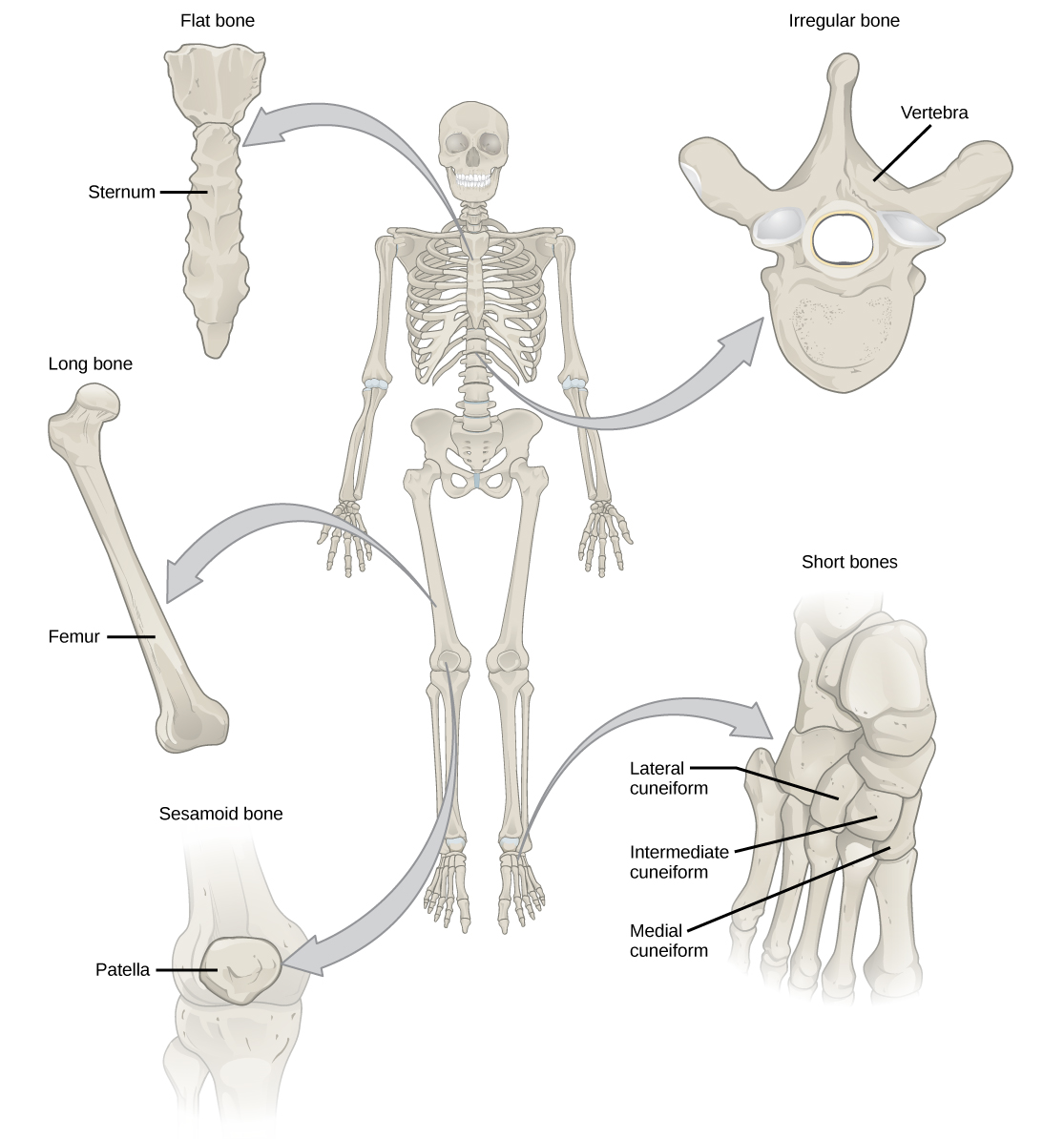
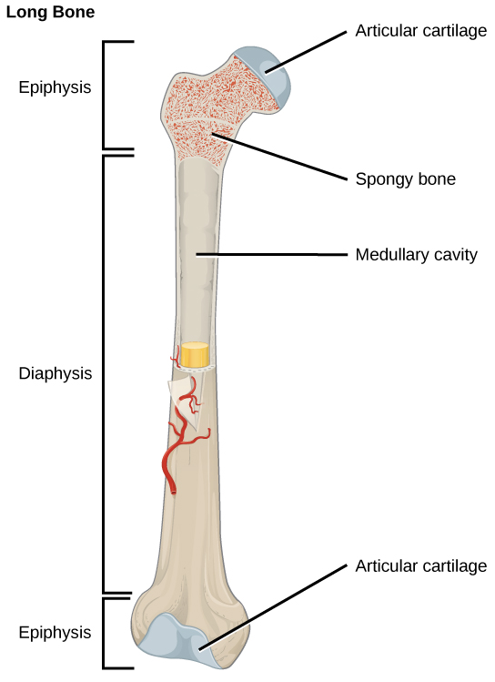
长骨比宽度长,有一根轴和两端。 diaphysis 或中心轴在骨髓腔中含有骨髓。 圆形末端,即骨髓,被关节软骨覆盖,并充满红骨髓,红骨髓会产生血细胞(图\(\PageIndex{2}\))。 大多数肢体骨骼都是长骨——例如,股骨、胫骨、尺骨和桡骨。 例外情况包括膝盖骨以及手腕和脚踝的骨头。
短骨头或长方体骨头是具有相同宽度和长度的骨骼,使它们具有立方体状的形状。 例如,手腕(腕骨)和脚踝(睑板)的骨头是短骨(图\(\PageIndex{1}\))。
扁平骨是指较薄且相对较宽的骨骼,用于需要广泛保护器官或需要宽阔的肌肉附着表面。 扁平骨的例子包括胸骨(胸骨)、肋骨、肩骨(肩 blade 骨)和头骨顶部(图\(\PageIndex{1}\))。
不规则骨头是具有复杂形状的骨头。 这些骨骼的表面可能很短、平坦、有缺口或有脊状。 不规则骨骼的例子包括椎骨、髋骨和几根头骨头。
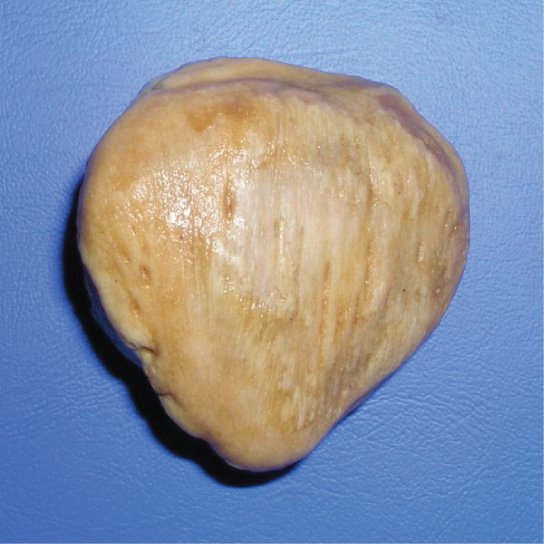
芝麻骨头是小而扁平的骨头,形状类似于芝麻种子。 髌骨是芝麻骨(图\(\PageIndex{3}\))。 芝麻骨发育在肌腱内,可能存在于膝盖、手和脚的关节附近。
缝合骨是小的、扁平的、形状不规则的骨头。 它们可能位于头骨的扁平骨头之间。 它们的数量、形状、大小和位置各不相同。
骨组织
骨骼被视为器官,因为它们含有各种类型的组织,例如血液、结缔组织、神经和骨组织。 骨细胞是骨组织的活细胞,形成骨骼的矿物质基质。 有两种类型的骨组织:紧凑型和海绵状。
紧凑型骨组织
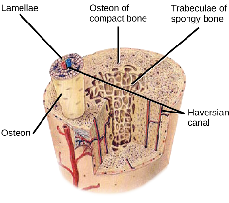
紧凑的骨头(或皮质骨)形成所有骨骼的坚硬外层,围绕着髓腔或骨髓。 它为骨骼提供保护和力量。 紧凑型骨组织由称为骨质或 Haversian 系统的单位组成。 骨质是圆柱形结构,含有矿物基质和由运送血液的 canaliculi 连接的活骨细胞。 它们平行于骨骼的长轴对齐。 每个 osteon 都由薄片组成,薄片是紧凑的基质层,围绕着一条名为哈弗西安运河的中央运河。 Haversian 运河(骨管)包含骨骼的血管和神经纤维(图\(\PageIndex{4}\))。 紧凑型骨组织中的骨沿着应力线朝相同的方向排列,有助于骨抵抗弯曲或骨折。 因此,紧凑型骨组织在仅向几个方向施加压力的骨骼区域很突出。
艺术连接
以下关于骨组织的陈述中哪一项是错误的?
- 紧凑的骨组织由圆柱形的骨组成,这些骨对齐使它们沿着骨骼的长度移动。
- 哈弗西安运河仅包含血管。
- Haversian 运河含有血管和神经纤维。
- 在骨头内部发现了海绵组织,在外部发现了紧凑的骨组织。
海绵状骨组织
紧凑的骨组织构成所有骨骼的外层,而海绵状骨或松质骨形成所有骨骼的内层。 海绵状骨组织不含构成紧凑型骨组织的骨素。 相反,它由小梁组成,小梁是以棒状或板状排列的薄片。 红骨髓存在于小梁之间。 该组织内的血管向骨细胞输送营养物质并清除废物。 股骨的红骨髓和其他大骨头(例如回肠)的内部形成血细胞。
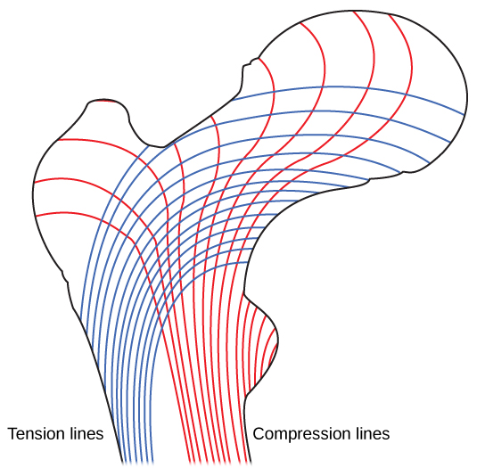
海绵状的骨头会降低骨的密度,并允许长骨的末端因对骨骼施加的压力而受到压缩。 海绵状骨头在没有严重压力的区域或应力来自多个方向的区域很突出。 骨骼的骨髓,例如股骨的脖子,受到来自多个方向的压力。 想象一下,将一张沉重的画框平放在地板上。 如果牙签垂直于地板和图片,你可以用牙签举起照片的一面。 现在钻一个洞然后把牙签塞进墙里把照片挂起来。 在这种情况下,牙签的作用是将图片的向下压力传递到墙上。 画面上的力量直接向下延伸到地板上,但牙签上的力量既是画线向下拉,又是墙洞底部向上推。 牙签会在墙上折断。
股骨的脖子是水平的,就像墙上的牙签一样。 身体的重量将其向下推到关节附近,但股骨的垂直透析将其推到另一端。 股骨的颈部必须足够强壮,才能将体重的向下力水平转移到股骨的垂直轴(图\(\PageIndex{5}\))。

骨骼中的细胞类型
骨由四种类型的细胞组成:成骨细胞、破骨细胞、骨细胞和骨祖细胞。 成骨细胞是负责骨形成的骨细胞。 成骨细胞合成并分泌骨组织和胶原纤维的细胞外基质的有机部分和无机部分。 成骨细胞被困在这些分泌物中,分化为活性较低的骨细胞。 破骨细胞是具有多达 50 个核的大型骨细胞。 它们通过释放溶解骨基质的溶酶体酶和酸来去除骨结构。 这些矿物质从骨骼释放到血液中,有助于调节体液中的钙浓度。 如果施加的应力发生了变化,骨骼也可以被吸收以进行重塑。 骨细胞是成熟的骨细胞,是骨结缔组织中的主要细胞;这些细胞无法分裂。 骨细胞通过回收骨基质中的矿物盐来维持正常的骨结构。 骨祖细胞是鳞状干细胞,分裂产生子细胞,分化为成骨细胞。 骨祖细胞在骨折修复中很重要。
骨骼的发育
骨化或成骨是成骨细胞形成骨骼的过程。 骨化不同于钙化过程;虽然钙化发生在骨化期间,但也可能发生在其他组织中。 骨化在胚胎受精大约六周后开始。 在此之前,胚胎骨骼完全由纤维膜和透明软骨组成。 由纤维膜发育的骨骼称为膜内骨化;由透明软骨发育的称为软骨内骨化。 骨骼的生长一直持续到大约 25 岁。 一生中骨骼的厚度都会增长,但是在25岁以后,骨化主要起到骨重塑和修复的作用。
膜内骨化
膜内骨化是纤维膜发育骨骼的过程。 它参与头骨、下颌骨和锁骨的扁平骨头的形成。 骨化始于间充质细胞形成未来骨骼的模板。 然后,它们在骨化中心分化为成骨细胞。 成骨细胞分泌细胞外基质并沉积钙,从而使基质变硬。 骨骼或类骨的非矿化部分继续在血管周围形成,形成海绵状的骨头。 基质中的结缔组织在胎儿中分化为红色骨髓。 海绵状的骨头在海绵状骨的表面被重塑成一层薄薄的紧凑型骨头。
软骨内骨化
软骨内骨化是从透明软骨中发育骨骼的过程。 除了头骨、下颌骨和锁骨的扁平骨外,身体的所有骨骼都是通过软骨内骨化形成的。
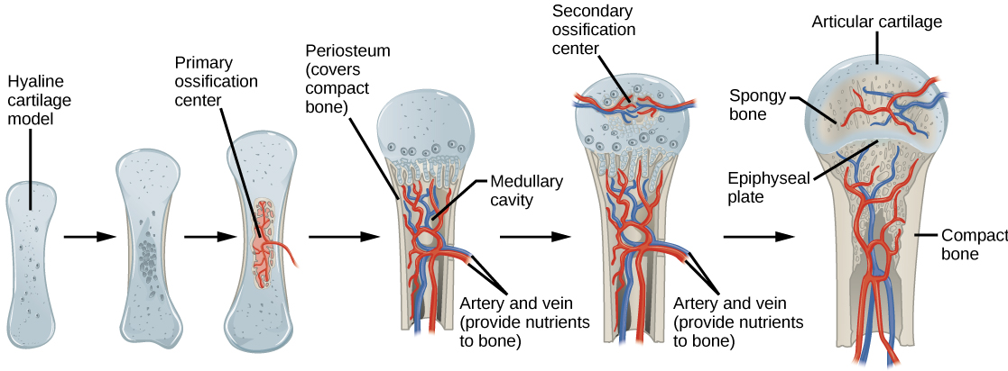
在长骨中,软骨细胞构成透明软骨透析的模板。 对复杂的发育信号作出反应,基质开始钙化。 这种钙化会阻止营养物质扩散到基质中,从而导致软骨细胞死亡并打开透析软骨中的空腔。 血管侵入蛀牙,成骨细胞和破骨细胞将钙化的软骨基质改造成海绵状的骨骼。 然后,破骨细胞分解一些海绵状的骨头,在透析中心形成骨髓或髓质腔。 密集、不规则的结缔组织在骨骼周围形成鞘层(骨膜)。 骨膜有助于将骨头附着到周围的组织、肌腱和韧带上。 随着骨髓软骨细胞的分裂,骨骼继续生长和伸长。
在产前骨发育的最后阶段,骨髓中心开始钙化。 当血管和成骨细胞进入骨髓并将透明软骨转化为海绵状骨时,在骨髓中形成继发骨化中心。 直到青春期,透明软骨一直存在于 e piphyseal plate(生长板),这是介于透析和骨髓之间的区域,是长骨纵向生长的原因(图\(\PageIndex{6}\)).
Growth of Bone
Long bones continue to lengthen, potentially until adolescence, through the addition of bone tissue at the epiphyseal plate. They also increase in width through appositional growth.
Lengthening of Long Bones
Chondrocytes on the epiphyseal side of the epiphyseal plate divide; one cell remains undifferentiated near the epiphysis, and one cell moves toward the diaphysis. The cells, which are pushed from the epiphysis, mature and are destroyed by calcification. This process replaces cartilage with bone on the diaphyseal side of the plate, resulting in a lengthening of the bone.
Long bones stop growing at around the age of 18 in females and the age of 21 in males in a process called epiphyseal plate closure. During this process, cartilage cells stop dividing and all of the cartilage is replaced by bone. The epiphyseal plate fades, leaving a structure called the epiphyseal line or epiphyseal remnant, and the epiphysis and diaphysis fuse.
Thickening of Long Bones
Appositional growth is the increase in the diameter of bones by the addition of bony tissue at the surface of bones. Osteoblasts at the bone surface secrete bone matrix, and osteoclasts on the inner surface break down bone. The osteoblasts differentiate into osteocytes. A balance between these two processes allows the bone to thicken without becoming too heavy.
Bone Remodeling and Repair
Bone renewal continues after birth into adulthood. Bone remodeling is the replacement of old bone tissue by new bone tissue. It involves the processes of bone deposition by osteoblasts and bone resorption by osteoclasts. Normal bone growth requires vitamins D, C, and A, plus minerals such as calcium, phosphorous, and magnesium. Hormones such as parathyroid hormone, growth hormone, and calcitonin are also required for proper bone growth and maintenance.
Bone turnover rates are quite high, with five to seven percent of bone mass being recycled every week. Differences in turnover rate exist in different areas of the skeleton and in different areas of a bone. For example, the bone in the head of the femur may be fully replaced every six months, whereas the bone along the shaft is altered much more slowly.
Bone remodeling allows bones to adapt to stresses by becoming thicker and stronger when subjected to stress. Bones that are not subject to normal stress, for example when a limb is in a cast, will begin to lose mass. A fractured or broken bone undergoes repair through four stages:
- Blood vessels in the broken bone tear and hemorrhage, resulting in the formation of clotted blood, or a hematoma, at the site of the break. The severed blood vessels at the broken ends of the bone are sealed by the clotting process, and bone cells that are deprived of nutrients begin to die.
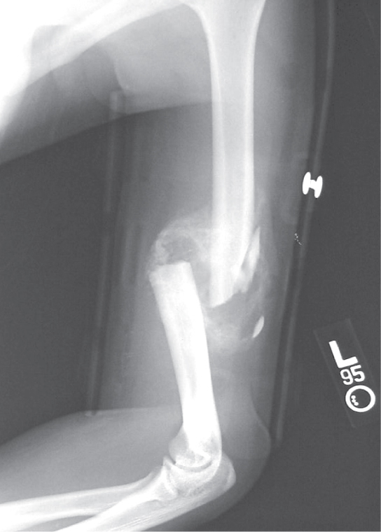
- Within days of the fracture, capillaries grow into the hematoma, and phagocytic cells begin to clear away the dead cells. Though fragments of the blood clot may remain, fibroblasts and osteoblasts enter the area and begin to reform bone. Fibroblasts produce collagen fibers that connect the broken bone ends, and osteoblasts start to form spongy bone. The repair tissue between the broken bone ends is called the fibrocartilaginous callus, as it is composed of both hyaline and fibrocartilage (Figure \(\PageIndex{7}\)). Some bone spicules may also appear at this point.
- The fibrocartilaginous callus is converted into a bony callus of spongy bone. It takes about two months for the broken bone ends to be firmly joined together after the fracture. This is similar to the endochondral formation of bone, as cartilage becomes ossified; osteoblasts, osteoclasts, and bone matrix are present.
- The bony callus is then remodelled by osteoclasts and osteoblasts, with excess material on the exterior of the bone and within the medullary cavity being removed. Compact bone is added to create bone tissue that is similar to the original, unbroken bone. This remodeling can take many months, and the bone may remain uneven for years.
Scientific Method Connection: Decalcification of Bones
Question: What effect does the removal of calcium and collagen have on bone structure?
Background: Conduct a literature search on the role of calcium and collagen in maintaining bone structure. Conduct a literature search on diseases in which bone structure is compromised.
Hypothesis: Develop a hypothesis that states predictions of the flexibility, strength, and mass of bones that have had the calcium and collagen components removed. Develop a hypothesis regarding the attempt to add calcium back to decalcified bones.
Test the hypothesis: Test the prediction by removing calcium from chicken bones by placing them in a jar of vinegar for seven days. Test the hypothesis regarding adding calcium back to decalcified bone by placing the decalcified chicken bones into a jar of water with calcium supplements added. Test the prediction by denaturing the collagen from the bones by baking them at 250°C for three hours.
Analyze the data: Create a table showing the changes in bone flexibility, strength, and mass in the three different environments.
Report the results: Under which conditions was the bone most flexible? Under which conditions was the bone the strongest?
Draw a conclusion: Did the results support or refute the hypothesis? How do the results observed in this experiment correspond to diseases that destroy bone tissue?
Summary
Bone, or osseous tissue, is connective tissue that includes specialized cells, mineral salts, and collagen fibers. The human skeleton can be divided into long bones, short bones, flat bones, and irregular bones. Compact bone tissue is composed of osteons and forms the external layer of all bones. Spongy bone tissue is composed of trabeculae and forms the inner part of all bones. Four types of cells compose bony tissue: osteocytes, osteoclasts, osteoprogenitor cells, and osteoblasts. Ossification is the process of bone formation by osteoblasts. Intramembranous ossification is the process of bone development from fibrous membranes. Endochondral ossification is the process of bone development from hyaline cartilage. Long bones lengthen as chondrocytes divide and secrete hyaline cartilage. Osteoblasts replace cartilage with bone. Appositional growth is the increase in the diameter of bones by the addition of bone tissue at the surface of bones. Bone remodeling involves the processes of bone deposition by osteoblasts and bone resorption by osteoclasts. Bone repair occurs in four stages and can take several months.
Art Exercise
Figure \(\PageIndex{4}\): Which of the following statements about bone tissue is false?
- Compact bone tissue is made of cylindrical osteons that are aligned such that they travel the length of the bone.
- Haversian canals contain blood vessels only.
- Haversian canals contain blood vessels and nerve fibers.
- Spongy tissue is found on the interior of the bone, and compact bone tissue is found on the exterior.
- Answer
-
B
Glossary
- appositional growth
- increase in the diameter of bones by the addition of bone tissue at the surface of bones
- bone
- (also, osseous tissue) connective tissue that constitutes the endoskeleton
- bone remodeling
- replacement of old bone tissue by new bone tissue
- calcification
- process of deposition of mineral salts in the collagen fiber matrix that crystallizes and hardens the tissue
- compact bone
- forms the hard external layer of all bones
- diaphysis
- central shaft of bone, contains bone marrow in a marrow cavity
- endochondral ossification
- process of bone development from hyaline cartilage
- epiphyseal plate
- region between the diaphysis and epiphysis that is responsible for the lengthwise growth of long bones
- epiphysis
- rounded end of bone, covered with articular cartilage and filled with red bone marrow, which produces blood cells
- flat bone
- thin and relatively broad bone found where extensive protection of organs is required or where broad surfaces of muscle attachment are required
- Haversian canal
- contains the bone’s blood vessels and nerve fibers
- intramembranous ossification
- process of bone development from fibrous membranes
- irregular bone
- bone with complex shapes; examples include vertebrae and hip bones
- lamella
- layer of compact tissue that surrounds a central canal called the Haversian canal
- long bone
- bone that is longer than wide, and has a shaft and two ends
- osteoblast
- bone cell responsible for bone formation
- osteoclast
- large bone cells with up to 50 nuclei, responsible for bone remodeling
- osteocyte
- mature bone cells and the main cell in bone tissue
- osseous tissue
- connective tissue that constitutes the endoskeleton
- ossification
- (also, osteogenesis) process of bone formation by osteoblasts
- osteon
- cylindrical structure aligned parallel to the long axis of the bone
- resorption
- process by which osteoclasts release minerals stored in bones
- sesamoid bone
- small, flat bone shaped like a sesame seed; develops inside tendons
- short bone
- bone that has the same width and length, giving it a cube-like shape
- spongy bone tissue
- forms the inner layer of all bones
- suture bone
- small, flat, irregularly shaped bone that forms between the flat bones of the cranium
- trabeculae
- lamellae that are arranged as rods or plates