
43.6: الإخصاب والتطور الجنيني المبكر
- Page ID
- 196293

المهارات اللازمة للتطوير
- ناقش كيفية حدوث الإخصاب
- اشرح كيف يتشكل الجنين من الزيجوت
- ناقش دور الانقسام والتذوق في تنمية الحيوان
إن العملية التي يتطور فيها الكائن الحي من زيجوت أحادي الخلية إلى كائن متعدد الخلايا معقدة ومنظمة جيدًا. تعتبر المراحل المبكرة من التطور الجنيني ضرورية أيضًا لضمان لياقة الكائن الحي.
التخصيب
الإخصاب، الموضح في الشكل\(\PageIndex{1}\) أ هو العملية التي تندمج فيها الأمشاج (البويضة والحيوانات المنوية) لتكوين زيجوت. تحتوي كل من البويضة والحيوانات المنوية على مجموعة واحدة من الكروموسومات. للتأكد من أن النسل يحتوي على مجموعة واحدة كاملة من الكروموسومات ثنائية الصبغيات، يجب أن يندمج حيوان منوي واحد فقط مع بويضة واحدة. في الثدييات، تتم حماية البويضة بطبقة من المصفوفة خارج الخلية تتكون أساسًا من بروتينات سكرية تسمى zona pellucida. عندما ترتبط الحيوانات المنوية بالمنطقة الشفافة، تحدث سلسلة من الأحداث البيوكيميائية، تسمى التفاعلات الأكروسومية. في الثدييات المشيمية، يحتوي الأكروسوم على إنزيمات هضمية تبدأ في تحلل مصفوفة البروتين السكري التي تحمي البويضة وتسمح لغشاء بلازما الحيوانات المنوية بالاندماج مع غشاء بلازما البيض، كما هو موضح في الشكل\(\PageIndex{1}\) (ب)، ويؤدي اندماج هذين الغشاءين إلى خلق فتحة من خلالها حيث يتم نقل نواة الحيوانات المنوية إلى البويضة. تتحلل الأغشية النووية للبويضة والحيوانات المنوية ويتكثف الجينومان أحادي الصبغيات لتشكيل جينوم ثنائي الصبغيات.
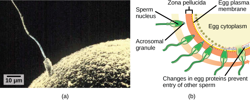
للتأكد من عدم قيام أكثر من حيوان منوي واحد بتخصيب البويضة، بمجرد حدوث التفاعلات الأكروسومية في موقع واحد من غشاء البويضة، تطلق البويضة البروتينات في مواقع أخرى لمنع الحيوانات المنوية الأخرى من الاندماج مع البويضة. إذا فشلت هذه الآلية، يمكن أن تندمج الحيوانات المنوية المتعددة مع البويضة، مما يؤدي إلى تعدد النطاف. الجنين الناتج غير قابل للحياة وراثيًا ويموت في غضون أيام قليلة.
مرحلة الانقسام والبلاستولا
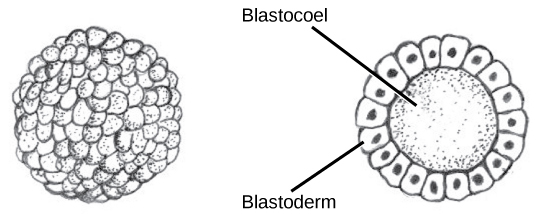
يبدأ تطور الكائنات الحية متعددة الخلايا من زيجوت أحادي الخلية، يخضع لانقسام سريع للخلايا لتشكيل الأريمة. تسمى الجولات السريعة والمتعددة من انقسام الخلايا بالانشقاق. يتم توضيح الانقسام في (الشكل\(\PageIndex{2}\) أ). بعد أن ينتج الانقسام أكثر من 100 خلية، يُطلق على الجنين اسم الأريمة. عادة ما تكون الأريمية عبارة عن طبقة كروية من الخلايا (الأريستوديرم) تحيط بتجويف مملوء بالسائل أو صفار البيض (الأريمية). تشكل الثدييات في هذه المرحلة بنية تسمى الكيسة الأريمية، وتتميز بكتلة خلية داخلية متميزة عن الأريمية المحيطة، كما هو موضح في الشكل\(\PageIndex{2}\) (ب)، وأثناء الانقسام، تنقسم الخلايا دون زيادة في الكتلة؛ أي أن زيجوت كبير أحادي الخلية ينقسم إلى عدة خلايا أصغر . تُسمى كل خلية داخل الأريمة بالبلاستومير.
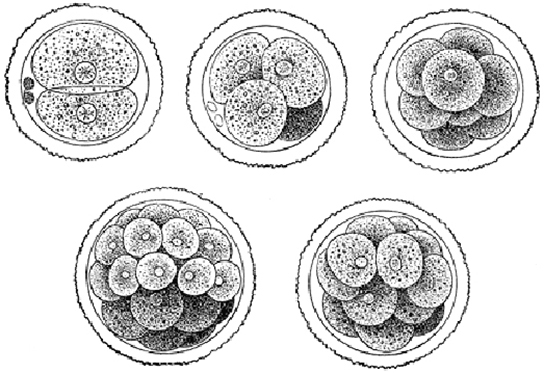
يمكن أن يحدث الانقسام بطريقتين: الانقسام الهولوبلاستيك (الكلي) أو الانقسام الميروبلاستي (الجزئي). يعتمد نوع الانقسام على كمية صفار البيض. في الثدييات المشيمية (بما في ذلك البشر) حيث يتم توفير التغذية من قبل جسم الأم، يحتوي البيض على كمية صغيرة جدًا من صفار البيض ويخضع لانشقاق هولوبلاستيك. الأنواع الأخرى، مثل الطيور، التي تحتوي على الكثير من صفار البيض لتغذية الجنين أثناء النمو، تخضع لانشقاق عضلي.
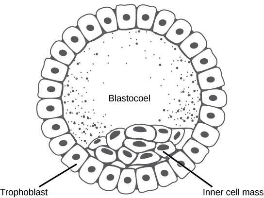
في الثدييات، تشكل الأريمة الكيسة الأريمية في المرحلة التالية من التطور. هنا تقوم الخلايا الموجودة في الأريمة بترتيب نفسها في طبقتين: كتلة الخلية الداخلية، وطبقة خارجية تسمى الأرومة الغاذية. تُعرف كتلة الخلية الداخلية أيضًا باسم الخلايا الجنينية وستستمر كتلة الخلايا هذه في تكوين الجنين. في هذه المرحلة من التطور، كما هو موضح في الشكل\(\PageIndex{3}\)، تتكون كتلة الخلية الداخلية من الخلايا الجذعية الجنينية التي ستتميز إلى أنواع الخلايا المختلفة التي يحتاجها الكائن الحي. سوف تساهم الأرومة الغاذية في المشيمة وتغذي الجنين.
رابط إلى التعلم

قم بزيارة مشروع الجنين البشري الافتراضي في موقع Endowment for Human Development للاطلاع على عرض تفاعلي يوضح مراحل تطور الجنين، بما في ذلك الصور المجهرية والصور الدوارة ثلاثية الأبعاد.
إلتهاب المعدة
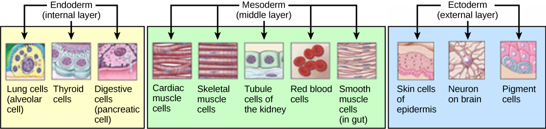
الأريمة النموذجية هي كرة من الخلايا. المرحلة التالية في التطور الجنيني هي تشكيل خطة الجسم. تقوم الخلايا في الأريمة بإعادة ترتيب نفسها مكانيًا لتشكيل ثلاث طبقات من الخلايا. هذه العملية تسمى المعدة. أثناء عملية المعدة، تطوى الأريمة على نفسها لتشكل الطبقات الثلاث من الخلايا. كل طبقة من هذه الطبقات تسمى طبقة جرثومية وكل طبقة جرثومية تفرق في أنظمة أعضاء مختلفة.
طبقات الجراثيم الثلاث، كما هو موضح في الشكل\(\PageIndex{4}\), are the endoderm, the ectoderm, and the mesoderm. The ectoderm gives rise to the nervous system and the epidermis. The mesoderm gives rise to the muscle cells and connective tissue in the body. The endoderm gives rise to columnar cells found in the digestive system and many internal organs.
Everyday Connection: Are Designer Babies in Our Future?
If you could prevent your child from getting a devastating genetic disease, would you do it? Would you select the sex of your child or select for their attractiveness, strength, or intelligence? How far would you go to maximize the possibility of resistance to disease? The genetic engineering of a human child, the production of "designer babies" with desirable phenotypic characteristics, was once a topic restricted to science fiction. This is the case no longer: science fiction is now overlapping into science fact. Many phenotypic choices for offspring are already available, with many more likely to be possible in the not too distant future. Which traits should be selected and how they should be selected are topics of much debate within the worldwide medical community. The ethical and moral line is not always clear or agreed upon, and some fear that modern reproductive technologies could lead to a new form of eugenics.
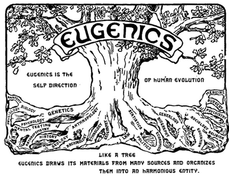
Eugenics is the use of information and technology from a variety of sources to improve the genetic makeup of the human race. The goal of creating genetically superior humans was quite prevalent (although controversial) in several countries during the early 20th century, but fell into disrepute when Nazi Germany developed an extensive eugenics program in the 1930's and 40's. As part of their program, the Nazis forcibly sterilized hundreds of thousands of the so-called "unfit" and killed tens of thousands of institutionally disabled people as part of a systematic program to develop a genetically superior race of Germans known as Aryans. Ever since, eugenic ideas have not been as publicly expressed, but there are still those who promote them.
Efforts have been made in the past to control traits in human children using donated sperm from men with desired traits. In fact, eugenicist Robert Klark Graham established a sperm bank in 1980 that included samples exclusively from donors with high IQs. The "genius" sperm bank failed to capture the public's imagination and the operation closed in 1999.
In more recent times, the procedure known as prenatal genetic diagnosis (PGD) has been developed. PGD involves the screening of human embryos as part of the process of in vitro fertilization, during which embryos are conceived and grown outside the mother's body for some period of time before they are implanted. The term PGD usually refers to both the diagnosis, selection, and the implantation of the selected embryos.
In the least controversial use of PGD, embryos are tested for the presence of alleles which cause genetic diseases such as sickle cell disease, muscular dystrophy, and hemophilia, in which a single disease-causing allele or pair of alleles has been identified. By excluding embryos containing these alleles from implantation into the mother, the disease is prevented, and the unused embryos are either donated to science or discarded. There are relatively few in the worldwide medical community that question the ethics of this type of procedure, which allows individuals scared to have children because of the alleles they carry to do so successfully. The major limitation to this procedure is its expense. Not usually covered by medical insurance and thus out of reach financially for most couples, only a very small percentage of all live births use such complicated methodologies. Yet, even in cases like these where the ethical issues may seem to be clear-cut, not everyone agrees with the morality of these types of procedures. For example, to those who take the position that human life begins at conception, the discarding of unused embryos, a necessary result of PGD, is unacceptable under any circumstances.
A murkier ethical situation is found in the selection of a child's sex, which is easily performed by PGD. Currently, countries such as Great Britain have banned the selection of a child's sex for reasons other than preventing sex-linked diseases. Other countries allow the procedure for "family balancing", based on the desire of some parents to have at least one child of each sex. Still others, including the United States, have taken a scattershot approach to regulating these practices, essentially leaving it to the individual practicing physician to decide which practices are acceptable and which are not.
Even murkier are rare instances of disabled parents, such as those with deafness or dwarfism, who select embryos via PGD to ensure that they share their disability. These parents usually cite many positive aspects of their disabilities and associated culture as reasons for their choice, which they see as their moral right. To others, to purposely cause a disability in a child violates the basic medical principle of Primum non nocere, "first, do no harm." This procedure, although not illegal in most countries, demonstrates the complexity of ethical issues associated with choosing genetic traits in offspring.
Where could this process lead? Will this technology become more affordable and how should it be used? With the ability of technology to progress rapidly and unpredictably, a lack of definitive guidelines for the use of reproductive technologies before they arise might make it difficult for legislators to keep pace once they are in fact realized, assuming the process needs any government regulation at all. Other bioethicists argue that we should only deal with technologies that exist now, and not in some uncertain future. They argue that these types of procedures will always be expensive and rare, so the fears of eugenics and "master" races are unfounded and overstated. The debate continues.
Summary
The early stages of embryonic development begin with fertilization. The process of fertilization is tightly controlled to ensure that only one sperm fuses with one egg. After fertilization, the zygote undergoes cleavage to form the blastula. The blastula, which in some species is a hollow ball of cells, undergoes a process called gastrulation, in which the three germ layers form. The ectoderm gives rise to the nervous system and the epidermal skin cells, the mesoderm gives rise to the muscle cells and connective tissue in the body, and the endoderm gives rise to columnar cells and internal organs.
Glossary
- acrosomal reaction
- series of biochemical reactions that the sperm uses to break through the zona pellucida
- blastocyst
- structure formed when cells in the mammalian blastula separate into an inner and outer layer
- gastrulation
- process in which the blastula folds over itself to form the three germ layers
- holoblastic
- complete cleavage; takes place in cells with a small amount of yolk
- inner cell mass
- inner layer of cells in the blastocyst
- meroblastic
- partial cleavage; takes place in cells with a large amount of yolk
- polyspermy
- condition in which one egg is fertilized by multiple sperm
- trophoblast
- outer layer of cells in the blastocyst
- zona pellucida
- protective layer of glycoproteins on the mammalian egg