
22.1: Viungo na Miundo ya Mfumo wa Kupumua
- Page ID
- 178525

Malengo ya kujifunza
- Andika orodha ya miundo inayounda mfumo wa kupumua
- Eleza jinsi mfumo wa kupumua unavyofanya oksijeni na CO 2
- Linganisha na kulinganisha kazi za njia ya kupumua ya juu na njia ya kupumua ya chini
Viungo vikuu vya mfumo wa kupumua hufanya kazi hasa kutoa oksijeni kwa tishu za mwili kwa kupumua kwa seli, kuondoa bidhaa taka dioksidi kaboni, na kusaidia kudumisha usawa wa asidi-msingi. Sehemu ya mfumo wa kupumua pia hutumiwa kwa kazi zisizo muhimu, kama vile kuhisi harufu, uzalishaji wa hotuba, na kwa kusisitiza, kama vile wakati wa kujifungua au kukohoa (Kielelezo\(\PageIndex{1}\)).
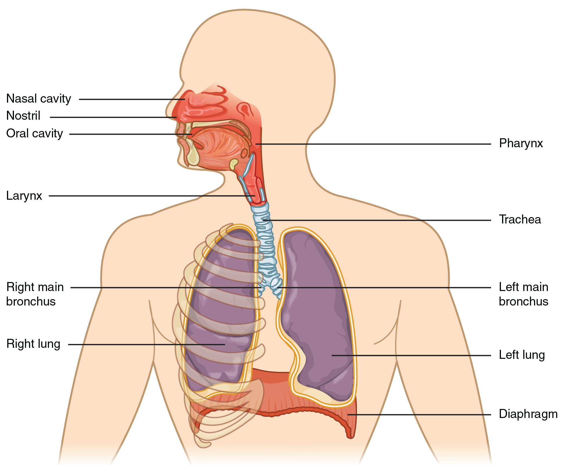
Kazi, mfumo wa kupumua unaweza kugawanywa katika eneo la uendeshaji na eneo la kupumua. Eneo la uendeshaji wa mfumo wa kupumua linajumuisha viungo na miundo isiyohusika moja kwa moja katika kubadilishana gesi. Kubadilishana gesi hutokea katika eneo la kupumua.
Kufanya Eneo
Kazi kuu za eneo la uendeshaji ni kutoa njia ya hewa inayoingia na inayoondoka, kuondoa uchafu na vimelea kutoka hewa inayoingia, na joto na humidify hewa inayoingia. Miundo kadhaa ndani ya eneo la uendeshaji hufanya kazi nyingine pia. Epithelium ya vifungu vya pua, kwa mfano, ni muhimu kwa kuhisi harufu, na epithelium ya bronchial ambayo mistari ya mapafu inaweza metabolize baadhi ya kansa za hewa.
Pua na Miundo yake iliyo karibu
Mlango mkubwa na kuondoka kwa mfumo wa kupumua ni kupitia pua. Wakati wa kujadili pua, ni muhimu kugawanya katika sehemu mbili kuu: pua ya nje, na cavity ya pua au pua ya ndani.
Pua ya nje ina miundo ya uso na mifupa ambayo husababisha kuonekana nje ya pua na kuchangia kazi zake nyingi (Kielelezo\(\PageIndex{2}\)). Mzizi ni kanda ya pua iliyo kati ya nyusi. Daraja ni sehemu ya pua inayounganisha mizizi kwa pua zote. Nasi ya dorsum ni urefu wa pua. Kilele ni ncha ya pua. Kwenye upande wowote wa kilele, puani huundwa na alae (umoja = ala). Ala ni muundo wa cartilaginous ambao huunda upande wa nyuma wa kila naris (wingi = nares), au ufunguzi wa pua. Philtrum ni uso wa concave unaounganisha kilele cha pua hadi mdomo wa juu.
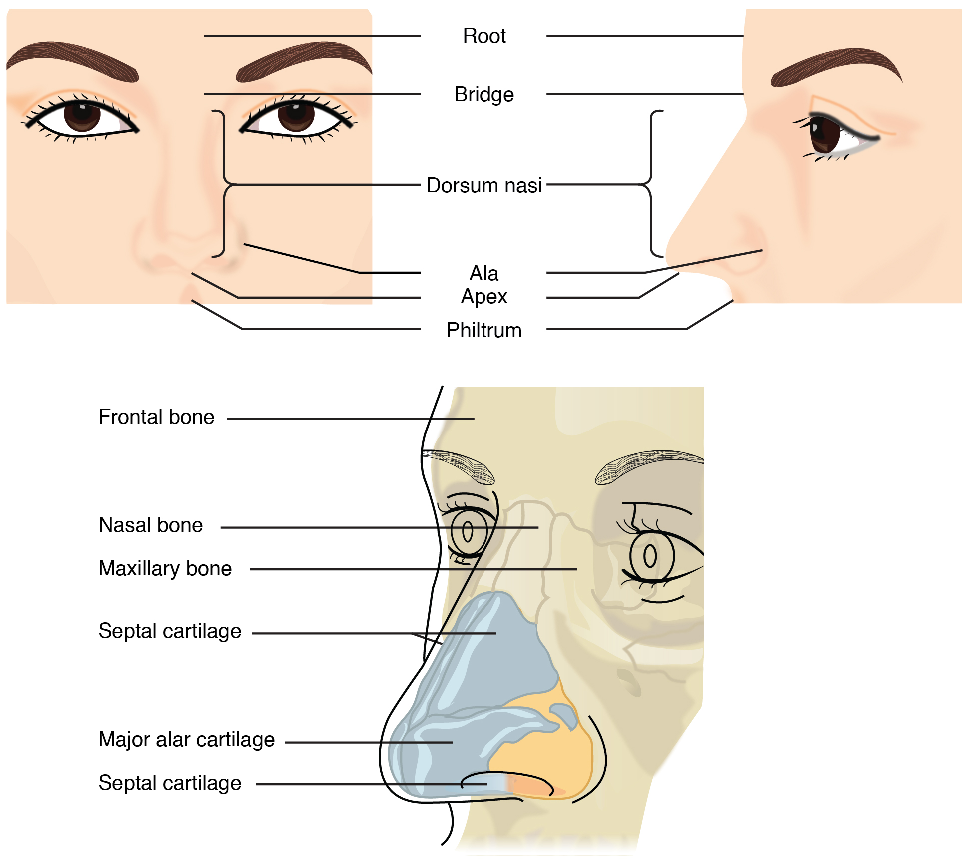
Chini ya ngozi nyembamba ya pua ni sifa zake za mifupa (angalia Mchoro, mfano wa chini). Wakati mizizi na daraja la pua linajumuisha mfupa, sehemu inayoendelea ya pua inajumuisha kamba. Matokeo yake, wakati wa kuangalia fuvu, pua haipo. Mfupa wa pua ni moja ya mifupa mawili yaliyo chini ya mizizi na daraja la pua. Mfupa wa pua unaelezea vizuri na mfupa wa mbele na baadaye na mifupa ya maxillary. Cartilage ya septal ni rahisi ya hyaline cartilage iliyounganishwa na mfupa wa pua, na kutengeneza nasi ya dorsum. Cartilage ya alar ina kilele cha pua; inazunguka naris.
Nares hufungua ndani ya cavity ya pua, ambayo imetenganishwa katika sehemu za kushoto na za kulia na septum ya pua (Kielelezo\(\PageIndex{3}\)). Pua septamu sumu anteriorly sehemu ya cartilage septal (sehemu rahisi unaweza kugusa vidole) na posteriorly perpendicular sahani ya mfupa ethmoid (fuvu mfupa iko tu nyuma ya mifupa ya pua) na nyembamba vomer mifupa (jina lake linamaanisha sura yake ya kulima). Kila ukuta wa mviringo wa cavity ya pua una makadirio matatu ya bony, inayoitwa conchae bora, katikati, na duni ya pua. Conchae duni ni mifupa tofauti, wakati conchae bora na ya kati ni sehemu ya mfupa wa ethmoid. Conchae hutumikia kuongeza eneo la uso wa cavity ya pua na kuharibu mtiririko wa hewa unapoingia pua, na kusababisha hewa kuinama pamoja na epithelium, ambako husafishwa na kuwaka. Conchae na meatuses pia huhifadhi maji na kuzuia maji mwilini ya epithelium ya pua kwa kunyakua maji wakati wa kutolea nje. Ghorofa ya cavity ya pua inajumuisha palate. Palate ngumu katika kanda ya anterior ya cavity ya pua inajumuisha mfupa. Palate laini katika sehemu ya nyuma ya cavity ya pua ina tishu za misuli. Air hutoka kwenye cavities ya pua kupitia nares ya ndani na huenda kwenye pharynx.

Mifupa kadhaa ambayo husaidia kuunda kuta za cavity ya pua ina nafasi zenye hewa zinazoitwa sinuses za paranasal, ambazo hutumikia joto na humidify hewa inayoingia. Sinuses zimewekwa na mucosa. Kila sinus paranasal inaitwa kwa mfupa wake unaohusishwa: sinus ya mbele, sinus maxillary, sinus sphenoidal, na sinus ethmoidal. Sinuses huzalisha kamasi na kupunguza uzito wa fuvu.
Nares na sehemu ya anterior ya cavity pua ni lined na kiwamboute, zenye tezi sebaceous na follicles nywele ambayo kutumika kuzuia kifungu cha uchafu kubwa, kama vile uchafu, kupitia cavity pua. Epithelium yenye ufanisi inayotumiwa kuchunguza harufu hupatikana zaidi katika cavity ya pua.
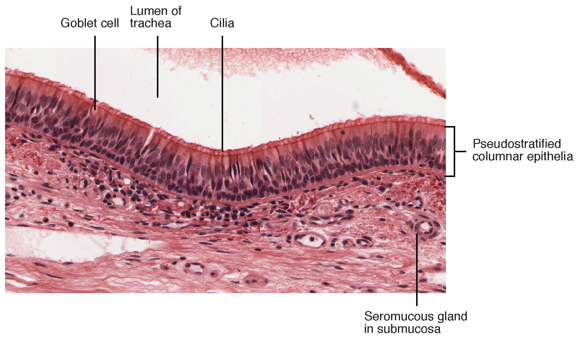
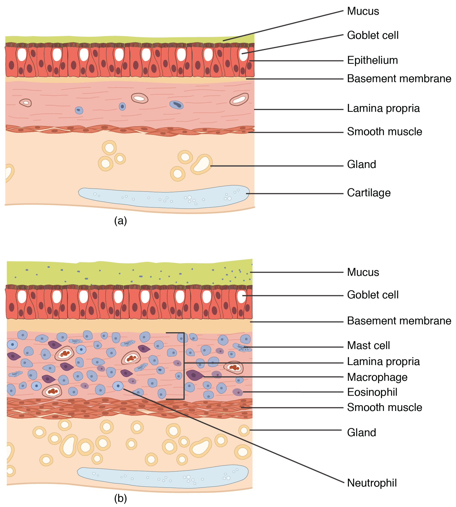
Conchae, meatuses, na sinuses paranasal ni lined na epithelium kupumua linajumuisha pseudostratified ciliated columnar epithelium (Kielelezo\(\PageIndex{4}\)). Epithelium ina seli za goblet, mojawapo ya seli za epithelial maalumu, za columnar zinazozalisha kamasi kwa mtego wa uchafu. Cilia ya epithelium ya kupumua husaidia kuondoa kamasi na uchafu kutoka kwenye cavity ya pua na mwendo wa kupiga mara kwa mara, vifaa vinavyojitokeza kuelekea koo ili kumeza. Kushangaza, hewa baridi hupunguza harakati za cilia, na kusababisha mkusanyiko wa kamasi ambayo inaweza kusababisha pua ya kukimbia wakati wa hali ya hewa ya baridi. Epithelium hii yenye unyevu hufanya kazi kwa joto na humidify hewa inayoingia. Capillaries iko chini ya epithelium ya pua hupunguza hewa kwa convection. Seli za serous na kamasi zinazozalisha pia hutoa enzyme ya lysozyme na protini inayoitwa defensins, ambayo ina mali ya antibacterial. Siri za kinga ambazo hupiga tishu zinazojumuisha kina kwa epithelium ya kupumua hutoa ulinzi wa ziada.

Pharynx
Pharynx ni tube inayotengenezwa na misuli ya mifupa na imefungwa na utando wa mucous unaoendelea na ule wa cavities ya pua (angalia Mchoro\(\PageIndex{3}\)). The pharynx is divided into three major regions: the nasopharynx, the oropharynx, and the laryngopharynx (Figure \(\PageIndex{5}\)).
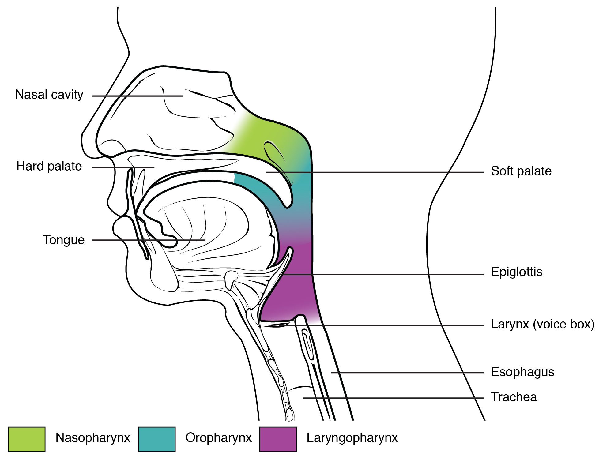
The nasopharynx is flanked by the conchae of the nasal cavity, and it serves only as an airway. At the top of the nasopharynx are the pharyngeal tonsils. A pharyngeal tonsil, also called an adenoid, is an aggregate of lymphoid reticular tissue similar to a lymph node that lies at the superior portion of the nasopharynx. The function of the pharyngeal tonsil is not well understood, but it contains a rich supply of lymphocytes and is covered with ciliated epithelium that traps and destroys invading pathogens that enter during inhalation. The pharyngeal tonsils are large in children, but interestingly, tend to regress with age and may even disappear. The uvula is a small bulbous, teardrop-shaped structure located at the apex of the soft palate. Both the uvula and soft palate move like a pendulum during swallowing, swinging upward to close off the nasopharynx to prevent ingested materials from entering the nasal cavity. In addition, auditory (Eustachian) tubes that connect to each middle ear cavity open into the nasopharynx. This connection is why colds often lead to ear infections.
The oropharynx is a passageway for both air and food. The oropharynx is bordered superiorly by the nasopharynx and anteriorly by the oral cavity. The fauces is the opening at the connection between the oral cavity and the oropharynx. As the nasopharynx becomes the oropharynx, the epithelium changes from pseudostratified ciliated columnar epithelium to stratified squamous epithelium. The oropharynx contains two distinct sets of tonsils, the palatine and lingual tonsils. A palatine tonsil is one of a pair of structures located laterally in the oropharynx in the area of the fauces. The lingual tonsil is located at the base of the tongue. Similar to the pharyngeal tonsil, the palatine and lingual tonsils are composed of lymphoid tissue, and trap and destroy pathogens entering the body through the oral or nasal cavities.
The laryngopharynx is inferior to the oropharynx and posterior to the larynx. It continues the route for ingested material and air until its inferior end, where the digestive and respiratory systems diverge. The stratified squamous epithelium of the oropharynx is continuous with the laryngopharynx. Anteriorly, the laryngopharynx opens into the larynx, whereas posteriorly, it enters the esophagus.
Larynx
The larynx is a cartilaginous structure inferior to the laryngopharynx that connects the pharynx to the trachea and helps regulate the volume of air that enters and leaves the lungs (Figure \(\PageIndex{6}\)). The structure of the larynx is formed by several pieces of cartilage. Three large cartilage pieces—the thyroid cartilage (anterior), epiglottis (superior), and cricoid cartilage (inferior)—form the major structure of the larynx. The thyroid cartilage is the largest piece of cartilage that makes up the larynx. The thyroid cartilage consists of the laryngeal prominence, or “Adam’s apple,” which tends to be more prominent in males. The thick cricoid cartilage forms a ring, with a wide posterior region and a thinner anterior region. Three smaller, paired cartilages—the arytenoids, corniculates, and cuneiforms—attach to the epiglottis and the vocal cords and muscle that help move the vocal cords to produce speech.
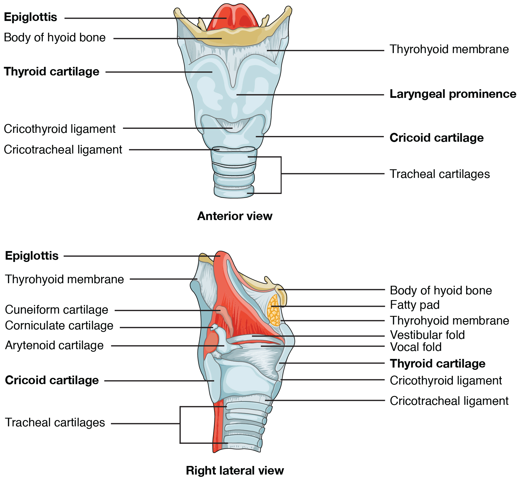
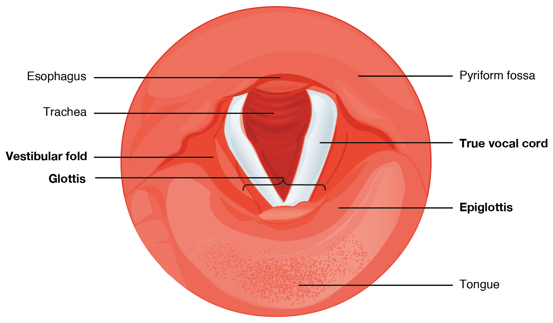
The epiglottis, attached to the thyroid cartilage, is a very flexible piece of elastic cartilage that covers the opening of the trachea (see Figure \(\PageIndex{3}\)). When in the “closed” position, the unattached end of the epiglottis rests on the glottis. The glottis is composed of the vestibular folds, the true vocal cords, and the space between these folds (Figure \(\PageIndex{7}\)). A vestibular fold, or false vocal cord, is one of a pair of folded sections of mucous membrane. A true vocal cord is one of the white, membranous folds attached by muscle to the thyroid and arytenoid cartilages of the larynx on their outer edges. The inner edges of the true vocal cords are free, allowing oscillation to produce sound. The size of the membranous folds of the true vocal cords differs between individuals, producing voices with different pitch ranges. Folds in males tend to be larger than those in females, which create a deeper voice. The act of swallowing causes the pharynx and larynx to lift upward, allowing the pharynx to expand and the epiglottis of the larynx to swing downward, closing the opening to the trachea. These movements produce a larger area for food to pass through, while preventing food and beverages from entering the trachea.
Continuous with the laryngopharynx, the superior portion of the larynx is lined with stratified squamous epithelium, transitioning into pseudostratified ciliated columnar epithelium that contains goblet cells. Similar to the nasal cavity and nasopharynx, this specialized epithelium produces mucus to trap debris and pathogens as they enter the trachea. The cilia beat the mucus upward towards the laryngopharynx, where it can be swallowed down the esophagus.
Trachea
The trachea (windpipe) extends from the larynx toward the lungs (Figure \(\PageIndex{8}\).a). The trachea is formed by 16 to 20 stacked, C-shaped pieces of hyaline cartilage that are connected by dense connective tissue. The trachealis muscle and elastic connective tissue together form the fibroelastic membrane, a flexible membrane that closes the posterior surface of the trachea, connecting the C-shaped cartilages. The fibroelastic membrane allows the trachea to stretch and expand slightly during inhalation and exhalation, whereas the rings of cartilage provide structural support and prevent the trachea from collapsing. In addition, the trachealis muscle can be contracted to force air through the trachea during exhalation. The trachea is lined with pseudostratified ciliated columnar epithelium, which is continuous with the larynx. The esophagus borders the trachea posteriorly.

Bronchial Tree
The trachea branches into the right and left primary bronchi at the carina. These bronchi are also lined by pseudostratified ciliated columnar epithelium containing mucus-producing goblet cells (Figure \(\PageIndex{8}\).b). The carina is a raised structure that contains specialized nervous tissue that induces violent coughing if a foreign body, such as food, is present. Rings of cartilage, similar to those of the trachea, support the structure of the bronchi and prevent their collapse. The primary bronchi enter the lungs at the hilum, a concave region where blood vessels, lymphatic vessels, and nerves also enter the lungs. The bronchi continue to branch into bronchial a tree. A bronchial tree (or respiratory tree) is the collective term used for these multiple-branched bronchi. The main function of the bronchi, like other conducting zone structures, is to provide a passageway for air to move into and out of each lung. In addition, the mucous membrane traps debris and pathogens.
A bronchiole branches from the tertiary bronchi. Bronchioles, which are about 1 mm in diameter, further branch until they become the tiny terminal bronchioles, which lead to the structures of gas exchange. There are more than 1000 terminal bronchioles in each lung. The muscular walls of the bronchioles do not contain cartilage like those of the bronchi. This muscular wall can change the size of the tubing to increase or decrease airflow through the tube.
Respiratory Zone
In contrast to the conducting zone, the respiratory zone includes structures that are directly involved in gas exchange. The respiratory zone begins where the terminal bronchioles join a respiratory bronchiole, the smallest type of bronchiole (Figure \(\PageIndex{9}\)), which then leads to an alveolar duct, opening into a cluster of alveoli.
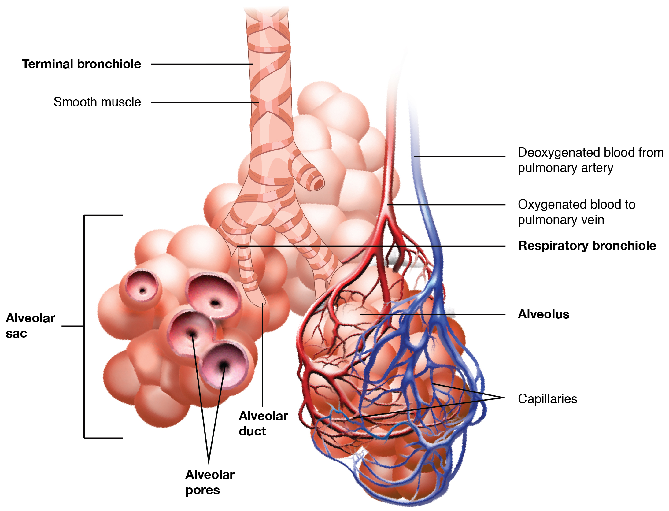
Alveoli
An alveolar duct is a tube composed of smooth muscle and connective tissue, which opens into a cluster of alveoli. An alveolus is one of the many small, grape-like sacs that are attached to the alveolar ducts.
An alveolar sac is a cluster of many individual alveoli that are responsible for gas exchange. An alveolus is approximately 200 μm in diameter with elastic walls that allow the alveolus to stretch during air intake, which greatly increases the surface area available for gas exchange. Alveoli are connected to their neighbors by alveolar pores, which help maintain equal air pressure throughout the alveoli and lung (Figure \(\PageIndex{10}\)).
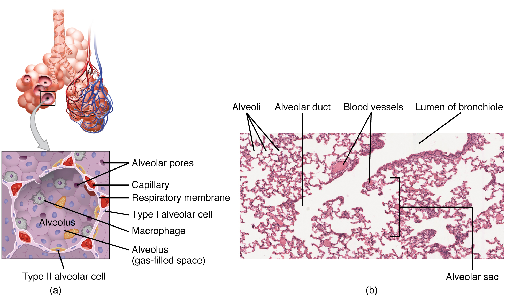
The alveolar wall consists of three major cell types: type I alveolar cells, type II alveolar cells, and alveolar macrophages. A type I alveolar cell is a squamous epithelial cell of the alveoli, which constitute up to 97 percent of the alveolar surface area. These cells are about 25 nm thick and are highly permeable to gases. A type II alveolar cell is interspersed among the type I cells and secretes pulmonary surfactant, a substance composed of phospholipids and proteins that reduces the surface tension of the alveoli. Roaming around the alveolar wall is the alveolar macrophage, a phagocytic cell of the immune system that removes debris and pathogens that have reached the alveoli.
The simple squamous epithelium formed by type I alveolar cells is attached to a thin, elastic basement membrane. This epithelium is extremely thin and borders the endothelial membrane of capillaries. Taken together, the alveoli and capillary membranes form a respiratory membrane that is approximately 0.5 mm thick. The respiratory membrane allows gases to cross by simple diffusion, allowing oxygen to be picked up by the blood for transport and CO2 to be released into the air of the alveoli.
Respiratory System: Asthma
Asthma is common condition that affects the lungs in both adults and children. Approximately 8.2 percent of adults (18.7 million) and 9.4 percent of children (7 million) in the United States suffer from asthma. In addition, asthma is the most frequent cause of hospitalization in children.
Asthma is a chronic disease characterized by inflammation and edema of the airway, and bronchospasms (that is, constriction of the bronchioles), which can inhibit air from entering the lungs. In addition, excessive mucus secretion can occur, which further contributes to airway occlusion (Figure \(\PageIndex{11}\)). Cells of the immune system, such as eosinophils and mononuclear cells, may also be involved in infiltrating the walls of the bronchi and bronchioles.
Bronchospasms occur periodically and lead to an “asthma attack.” An attack may be triggered by environmental factors such as dust, pollen, pet hair, or dander, changes in the weather, mold, tobacco smoke, and respiratory infections, or by exercise and stress.
Symptoms of an asthma attack involve coughing, shortness of breath, wheezing, and tightness of the chest. Symptoms of a severe asthma attack that requires immediate medical attention would include difficulty breathing that results in blue (cyanotic) lips or face, confusion, drowsiness, a rapid pulse, sweating, and severe anxiety. The severity of the condition, frequency of attacks, and identified triggers influence the type of medication that an individual may require. Longer-term treatments are used for those with more severe asthma. Short-term, fast-acting drugs that are used to treat an asthma attack are typically administered via an inhaler. For young children or individuals who have difficulty using an inhaler, asthma medications can be administered via a nebulizer.
In many cases, the underlying cause of the condition is unknown. However, recent research has demonstrated that certain viruses, such as human rhinovirus C (HRVC), and the bacteria Mycoplasma pneumoniae and Chlamydia pneumoniae that are contracted in infancy or early childhood, may contribute to the development of many cases of asthma.

Tembelea tovuti hii ili ujifunze zaidi kuhusu kinachotokea wakati wa mashambulizi ya pumu. Mabadiliko matatu yanayotokea ndani ya hewa wakati wa mashambulizi ya pumu ni nini?
Sura ya Mapitio
Mfumo wa kupumua ni wajibu wa kupata oksijeni na kuondokana na dioksidi kaboni, na kusaidia katika uzalishaji wa hotuba na kuhisi harufu. Kutokana na mtazamo wa kazi, mfumo wa kupumua unaweza kugawanywa katika maeneo mawili makubwa: eneo la uendeshaji na eneo la kupumua. Eneo la uendeshaji lina miundo yote ambayo hutoa njia za hewa kusafiri ndani na nje ya mapafu: cavity ya pua, pharynx, trachea, bronchi, na bronchioles nyingi. Vifungu vya pua vina vyenye conchae na nyama ambazo hupanua eneo la uso wa cavity, ambayo husaidia joto na humidify hewa inayoingia, huku kuondoa uchafu na vimelea. Pharynx inajumuisha sehemu tatu kuu: nasopharynx, ambayo inaendelea na cavity ya pua; oropharynx, ambayo inapakana na nasopharynx na cavity ya mdomo; na laryngopharynx, ambayo inapakana na oropharynx, trachea, na umio. Eneo la kupumua linajumuisha miundo ya mapafu ambayo inahusika moja kwa moja katika kubadilishana gesi: bronchioles ya terminal na alveoli.
Uchimbaji wa eneo la uendeshaji linajumuisha zaidi ya epithelium ya safu ya pseudostratified ciliated na seli za goblet. Mitego ya kamasi ya vimelea na uchafu, wakati kumpiga cilia husababisha kamasi kuelekea koo, ambako imemeza. Kama bronchioles kuwa ndogo na ndogo, na karibu na alveoli, epithelium thins na ni rahisi squamous epithelium katika alveoli. Endothelium ya capillaries zinazozunguka, pamoja na epithelium ya alveolar, hufanya utando wa kupumua. Hii ni kizuizi cha damu-hewa kwa njia ambayo kubadilishana gesi hutokea kwa kutenganishwa rahisi.
Maswali ya Link Interactive
Tembelea tovuti hii ili ujifunze zaidi kuhusu kinachotokea wakati wa mashambulizi ya pumu. Mabadiliko matatu yanayotokea ndani ya hewa wakati wa mashambulizi ya pumu ni nini?
Jibu: Kuvimba na uzalishaji wa kamasi nyembamba; kikwazo cha misuli ya hewa, au bronchospasm; na kuongezeka kwa unyeti kwa allergens.
Mapitio ya Maswali
Swali: Ni ipi kati ya miundo ya anatomical ifuatayo si sehemu ya eneo la uendeshaji?
A. pharynx
B. cavity ya pua
C. alveoli
D. bronchi
Jibu: C
Swali: Kazi ya conchae katika cavity ya pua ni nini?
A. kuongeza eneo la uso
B. gesi za kubadilishana
C. kudumisha mvutano uso
D. kudumisha shinikizo la hewa
Jibu: A
Swali: Fauces huunganisha ni ipi ya miundo ifuatayo kwa oropharynx?
A. nasopharynx
B. laryngopharynx
C. cavity ya pua
D. cavity ya mdomo
Jibu: D
Swali: Ni ipi kati ya yafuatayo ni vipengele vya miundo ya trachea?
A. cartilage ya umbo C
B. nyuzi za misuli laini
C. cilia
D. yote ya hapo juu
Jibu: A
Swali: Ni ipi kati ya miundo ifuatayo sio sehemu ya mti wa bronchial?
A. alveoli
B. bronchi
C. bronchioles mwisho
D. bronchioles kupumua
Jibu: C
Swali: Ni jukumu gani la macrophages ya alveolar?
A. ili kuzuia surfactant ya pulmona
B. ili kuzuia protini za antimicrobial
C. kuondoa vimelea na uchafu
D. kuwezesha kubadilishana gesi
Jibu: C
Maswali muhimu ya kufikiri
Swali: Eleza mikoa mitatu ya pharynx na kazi zao.
A. pharynx ina mikoa mitatu kuu. Mkoa wa kwanza ni nasopharynx, ambayo imeshikamana na cavity ya pua ya nyuma na inafanya kazi kama barabara ya hewa. Mkoa wa pili ni oropharynx, ambayo inaendelea na nasopharynx na imeshikamana na cavity ya mdomo kwenye fauces. Laryngopharynx imeshikamana na oropharynx na mkojo na trachea. Wote oropharynx na laryngopharynx ni njia za hewa na chakula na vinywaji.
Swali: Ikiwa mtu anaendelea kuumia kwa epiglottis, itakuwa matokeo gani ya kisaikolojia?
A. epiglottis ni kanda ya larynx ambayo ni muhimu wakati wa kumeza chakula au vinywaji. Kama mtu anavyomeza, pharynx huenda juu na epiglottis inafunga juu ya trachea, kuzuia chakula au kunywa kuingia kwenye trachea. Ikiwa epiglottis ya mtu ilijeruhiwa, utaratibu huu ungeharibika. Matokeo yake, mtu anaweza kuwa na shida na chakula au vinywaji kuingia kwenye trachea, na labda, mapafu. Baada ya muda, hii inaweza kusababisha maambukizi kama vile pneumonia kuingia.
Swali: Linganisha na kulinganisha maeneo ya uendeshaji na kupumua.
A. ukanda wa uendeshaji wa mfumo wa kupumua ni pamoja na viungo na miundo ambayo si moja kwa moja kushiriki katika kubadilishana gesi, lakini kutekeleza majukumu mengine kama vile kutoa njia ya hewa, utego na kuondoa uchafu na vimelea, na joto na humidifying hewa zinazoingia. Miundo kama hiyo ni pamoja na cavity ya pua, pharynx, larynx, trachea, na wengi wa mti wa bronchial. Eneo la kupumua linajumuisha viungo vyote na miundo inayohusika moja kwa moja katika kubadilishana gesi, ikiwa ni pamoja na bronchioles ya kupumua, ducts ya alveolar, na alveoli.
Marejeo
Bizzintino J, Lee WM, Leing IA, Vang F, Pappas T, Zhang G, Martin AC, Khoo SK, Cox DW, Geelhoed GC, nk. Chama kati ya rhinovirus C ya binadamu na ukali wa pumu kali kwa watoto. Eur Respir J [internet]. 2010 [alitoa mfano 2013 Machi 22]; 37 (5) :1037—1042. Inapatikana kutoka: erj.ersjournals.com/gca? submi..% 2F1037 & allch =
Kumar V, Ramzi S, Robbins SL. Robbins Basic Patholojia. 7 ed. Philadelphia (PA): Elsevier Ltd; 2005.
Martin RJ, Kraft M, Chu HW, Berns, EA, Cassell GH. Uhusiano kati ya pumu sugu na maambukizi ya muda mrefu. J Allergy Clin Immunol [Internet]. 2001 [alitoa mfano 2013 Machi 22]; 107 (4) :595-601. Inapatikana kutoka: erj.ersjournals.com/gca? submi..% 2F1037 & allch =
faharasa
- ala
- (wingi = alae) ndogo, muundo wa moto wa pua ambayo huunda upande wa nyuma wa nares
- alar cartilage
- cartilage ambayo inasaidia kilele cha pua na husaidia kuunda nares; imeunganishwa na cartilage ya septal na tishu zinazojumuisha za alae
- duct ya alveolar
- tube ndogo inayoongoza kutoka bronchiole ya terminal hadi bronchiole ya kupumua na ni hatua ya kushikamana kwa alveoli
- macrophage ya alveolar
- mfumo wa kinga ya seli ya alveolus kwamba kuondosha uchafu na vimelea
- pore ya alveolar
- ufunguzi ambayo inaruhusu airflow kati ya alveoli jirani
- kifuko cha alveolar
- nguzo ya alveoli
- alveolus
- ndogo, zabibu kama kifuko kwamba hufanya gesi kubadilishana katika mapafu
- kilele
- ncha ya pua ya nje
- mti wa bronchial
- jina la pamoja kwa matawi mengi ya bronchi na bronchioles ya mfumo wa kupumua
- daraja
- sehemu ya pua ya nje ambayo iko katika eneo la mifupa ya pua
- bronchiole
- tawi la bronchi ambayo ni 1 mm au chini ya kipenyo na kusitisha katika sacs alveolar
- bronchus
- tube kushikamana na trachea kwamba matawi katika matawi mengi na hutoa njia ya hewa kuingia na kuondoka mapafu
- eneo la kuendesha
- kanda ya mfumo wa kupumua ambayo ni pamoja na viungo na miundo ambayo hutoa njia za hewa na sio moja kwa moja kushiriki katika kubadilishana gesi
- cricoid cartilage
- sehemu ya larynx linajumuisha pete ya cartilage na mkoa mkubwa wa posterior na mkoa mwembamba wa anterior; masharti ya mkojo
- dorsum nasi
- sehemu ya kati ya pua ya nje inayounganisha daraja hadi kilele na inasaidiwa na mfupa wa pua
- epiglottis
- kipande cha jani cha kamba ya elastic ambayo ni sehemu ya larynx ambayo inakuja kufunga trachea wakati wa kumeza
- pua ya nje
- kanda ya pua inayoonekana kwa urahisi kwa wengine
- mifereji
- sehemu ya cavity posterior mdomo inayounganisha cavity mdomo kwa oropharynx
- fibroelastic utando
- utando maalumu unaounganisha mwisho wa cartilage ya sura ya C katika trachea; ina nyuzi za misuli nyembamba
- glottis
- kufungua kati ya mikunjo ya sauti ambayo hewa hupita wakati wa kuzalisha hotuba
- umaarufu wa laryngeal
- kanda ambapo mbili lamina ya cartilage tezi kujiunga, na kutengeneza protrusion inayojulikana kama “apple Adamu”
- laryngopharynx
- sehemu ya pharynx imepakana na oropharynx superiorly na umio na trachea inferiorly; hutumika kama njia ya hewa na chakula
- zoloto
- muundo wa cartilaginous unaozalisha sauti, huzuia chakula na vinywaji kuingia kwenye trachea, na inasimamia kiasi cha hewa kinachoingia na kuacha mapafu
- lingual tonsil
- tishu za lymphoid ziko chini ya ulimi
- nyama
- moja ya vifungo vitatu (bora, katikati, na duni) katika cavity ya pua iliyounganishwa na conchae ambayo huongeza eneo la uso wa cavity ya pua
- naris
- (wingi = nares) ufunguzi wa pua
- mfupa wa pua
- mfupa wa fuvu ambalo liko chini ya mizizi na daraja la pua na linaunganishwa na mifupa ya mbele na maxillary
- septum ya pua
- ukuta linajumuisha mfupa na cartilage kwamba hutenganisha cavities kushoto na kulia pua
- nasopharynx
- sehemu ya koo flanked na conchae na oropharynx ambayo hutumika kama airway
- oropharynx
- sehemu ya pharynx iliyozunguka na nasopharynx, cavity ya mdomo, na laryngopharynx ambayo ni njia ya hewa na chakula
- tonsil ya palatine
- moja ya miundo ya paired linajumuisha tishu za lymphoid ziko anterior kwa uvula kwenye paa la ismus ya fauces
- sinus paranasal
- moja ya cavities ndani ya fuvu iliyounganishwa na conchae ambayo hutumikia joto na humidify hewa inayoingia, kuzalisha kamasi, na kupunguza uzito wa fuvu; lina dhambi za mbele, maxillary, sphenoidal, na ethmoidal
- tonsil ya koromeo
- muundo linajumuisha tishu lymphoid iko katika nasopharynx
- koromeo
- kanda ya ukanda wa uendeshaji ambao huunda tube ya misuli ya mifupa iliyowekwa na epithelium ya kupumua; iko kati ya conchae ya pua na mimba na trachea
- philtrum
- concave uso wa uso unaounganisha kilele cha pua kwa mdomo wa juu
- surfactant ya mapafu
- dutu linajumuisha phospholipids na protini ambazo hupunguza mvutano wa uso wa alveoli; iliyofanywa na seli za aina ya II
- bronchiole ya kupumua
- aina maalum ya bronchiole inayoongoza kwa sacs ya alveolar
- epithelium kupumua
- ciliated bitana ya sehemu kubwa ya ukanda conductive kwamba ni maalumu kuondoa uchafu na vimelea, na kuzalisha kamasi
- utando wa kupumua
- ukuta wa alveolar na capillary pamoja, ambayo huunda kizuizi cha hewa-damu ambacho kinawezesha ugawanyiko rahisi wa gesi
- eneo la kupumua
- ni pamoja na miundo ya mfumo wa kupumua ambayo ni moja kwa moja kushiriki katika kubadilishana gesi
- mzizi
- kanda ya pua ya nje kati ya nyusi
- tezi cartilage
- kubwa kipande cha cartilage kwamba hufanya juu ya larynx na lina lamina mbili
- koo
- tube linajumuisha pete za cartilaginous na tishu zinazounga mkono zinazounganisha bronchi ya mapafu na larynx; hutoa njia ya hewa kuingia na kuondoka mapafu
- misuli ya trachealis
- misuli ya laini iko kwenye membrane ya fibroelastic ya trachea
- kamba ya kweli ya sauti
- moja ya jozi ya membrane iliyopigwa, nyeupe ambayo ina makali ya ndani ya bure ambayo hupunguza kama hewa inapita ili kuzalisha sauti
- aina mimi kiini cha alveolar
- seli za epithelial za squamous ambazo ni aina kuu ya seli katika ukuta wa alveolar; yenye kutosha kwa gesi
- aina ya II ya seli ya alveolar
- seli za epithelial za cuboidal ambazo ni aina ndogo ya seli katika ukuta wa alveolar; secrete surfactant ya pulmona
- pindo la nguo
- sehemu ya mkoa uliowekwa wa glottis linajumuisha utando wa mucous; inasaidia epiglottis wakati wa kumeza