
4.1: 研究细胞
- Page ID
- 202609

培养技能
- 描述细胞在生物体中的作用
- 比较和对比光学显微镜和电子显微镜
- 总结细胞理论
细胞是生物的最小单位。 生物无论是由一个细胞(如细菌)还是由多个细胞(如人类)组成,都被称为生物体。 因此,细胞是所有生物的基本组成部分。
几个相互连接并发挥共同功能的同一种细胞形成组织,几个组织结合形成一个器官(你的胃、心脏或大脑),几个器官组成一个器官系统(例如消化系统、循环系统或神经系统)。 几个共同起作用的系统形成一个有机体(比如人类)。 在这里,我们将研究细胞的结构和功能。
有许多类型的细胞,都分为两大类之一:原核细胞和真核细胞。 例如,动植物细胞都被归类为真核细胞,而细菌细胞被归类为原核细胞。 在讨论确定细胞是原核还是真核生物的标准之前,让我们先来看看生物学家是如何研究细胞的。
显微镜
细胞的大小各不相同。 除了少数例外,肉眼无法看到单个细胞,因此科学家使用显微镜(micro-= “小”;-scope = “看”)来研究它们。 显微镜是一种放大物体的仪器。 大多数细胞照片都是用显微镜拍摄的,这些图像也可以称为显微照片。
显微镜镜头的光学元件会改变用户看到的图像的方向。 通过显微镜观察时,在显微镜载玻片上右侧朝上且朝右的标本将出现颠倒和朝左,反之亦然。 同样,如果在透过显微镜观察时向左移动幻灯片,它似乎会向右移动,如果向下移动,则似乎向上移动。 之所以出现这种情况,是因为显微镜使用两组镜头来放大图像。 由于光线穿过镜头的方式,这种由两个镜头组成的系统会产生倒置图像(双筒望远镜或解剖显微镜的工作方式类似,但包括一个额外的放大系统,使最终图像看起来像直立)。
光学显微镜
为了让您了解细胞的大小,典型的人类红细胞直径约为八百万分之一米或八微米(缩写为八微米);针头的直径约为千分之二米(两毫米)。 这意味着大约 250 个红细胞可以装在别针的头上。
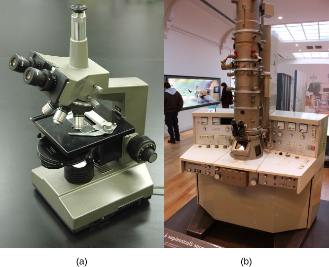
大多数学生显微镜被归类为光学显微镜(图\(\PageIndex{1}\) a)。 可见光通过并弯曲穿过镜头系统,使用户能够看到标本。 光学显微镜有利于观察活生物体,但由于单个细胞通常是透明的,因此除非用特殊的污渍着色,否则它们的成分是无法区分的。 但是,染色通常会杀死细胞。
本科院校实验室常用的光学显微镜最多可放大约 400 倍。 显微镜中两个重要的参数是放大倍率和分辨率。 放大是放大物体外观的过程。 分辨能力是显微镜能够将两个相邻结构分开分开:分辨率越高,图像的清晰度和细节就越好。 当使用油浸透镜研究小物体时,放大倍率通常会增加到 1,000 倍。 为了更好地了解细胞结构和功能,科学家通常使用电子显微镜。
电子显微镜
与光学显微镜相比,电子显微镜(图\(\PageIndex{1}\) b)使用电子束而不是光束。 这不仅允许更高的放大倍率,从而获得更多的细节(图\(\PageIndex{2}\)),而且还提供了更高的分辨率。 使用电子显微镜制备样本以供观察的方法会杀死标本。 电子具有短波长(比光子短),在真空中运动效果最好,因此无法用电子显微镜观察活细胞。
在扫描电子显微镜中,一束电子在细胞表面来回移动,形成细胞表面特征的细节。 在透射电子显微镜中,电子束穿透细胞并提供细胞内部结构的细节。 正如你可能想象的那样,电子显微镜比光学显微镜体积更大、更昂贵。

细胞理论
我们今天使用的显微镜比1700年代使用的显微镜要复杂得多,安东尼·范·勒文胡克是一位荷兰店主,在制作镜片方面有很高的技巧。 尽管他现在古老的镜片存在局限性,但范·勒文胡克还是观察了原生物(一种单细胞生物)和精子的运动,他统称为 “动物”。
在 1665 年出版的一本名为《Micrographia》的出版物中,实验科学家罗伯特·胡克为他在透镜观察软木组织时观察到的盒状结构创造了 “细胞” 一词。 在 16 世纪 70 年代,van Leeuwenhoek 发现了细菌和原生动物。 后来,镜头、显微镜结构和染色技术的进步使其他科学家能够看到细胞内的某些成分。
到19世纪30年代末,植物学家马蒂亚斯·施莱登和动物学家西奥多·施万正在研究组织并提出了统一的细胞理论,该理论指出,所有生物都由一个或多个细胞组成,细胞是生命的基本单位,新细胞来自现有细胞。 鲁道夫·维尔乔后来为这一理论做出了重要贡献。
职业联系:细胞技术专家
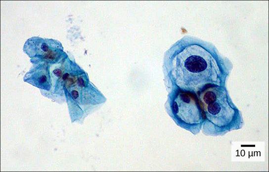
你听说过一种叫做子宫颈抹片检查的医学检查吗(图\(\PageIndex{3}\))? In this test, a doctor takes a small sample of cells from the uterine cervix of a patient and sends it to a medical lab where a cytotechnologist stains the cells and examines them for any changes that could indicate cervical cancer or a microbial infection.
Cytotechnologists (cyto- = “cell”) are professionals who study cells via microscopic examinations and other laboratory tests. They are trained to determine which cellular changes are within normal limits and which are abnormal. Their focus is not limited to cervical cells; they study cellular specimens that come from all organs. When they notice abnormalities, they consult a pathologist, who is a medical doctor who can make a clinical diagnosis.
Cytotechnologists play a vital role in saving people’s lives. When abnormalities are discovered early, a patient’s treatment can begin sooner, which usually increases the chances of a successful outcome.
Summary
A cell is the smallest unit of life. Most cells are so tiny that they cannot be seen with the naked eye. Therefore, scientists use microscopes to study cells. Electron microscopes provide higher magnification, higher resolution, and more detail than light microscopes. The unified cell theory states that all organisms are composed of one or more cells, the cell is the basic unit of life, and new cells arise from existing cells.
Glossary
- cell theory
- see unified cell theory
- electron microscope
- an instrument that magnifies an object using a beam of electrons passed and bent through a lens system to visualize a specimen
- light microscope
- an instrument that magnifies an object using a beam visible light passed and bent through a lens system to visualize a specimen
- microscope
- an instrument that magnifies an object
- unified cell theory
- a biological concept that states that all organisms are composed of one or more cells; the cell is the basic unit of life; and new cells arise from existing cells