
38.2: العظام
- Page ID
- 196153

مهارات التطوير
- صنف الأنواع المختلفة من العظام في الهيكل العظمي
- شرح دور أنواع الخلايا المختلفة في العظام
- شرح كيفية تشكل العظام أثناء النمو
العظام، أو الأنسجة العظمية، هي نسيج ضام يشكل الهيكل الداخلي. يحتوي على خلايا متخصصة ومصفوفة من الأملاح المعدنية وألياف الكولاجين.
تشمل الأملاح المعدنية بشكل أساسي هيدروكسيباتيت، وهو معدن يتكون من فوسفات الكالسيوم. التكلس هو عملية ترسب الأملاح المعدنية على مصفوفة ألياف الكولاجين التي تبلور وتصلب الأنسجة. تحدث عملية التكلس فقط في وجود ألياف الكولاجين.
يتم تصنيف عظام الهيكل العظمي البشري حسب شكلها: العظام الطويلة والعظام القصيرة والعظام المسطحة والعظام الخيطية والعظام السمسوية والعظام غير المنتظمة (الشكل\(\PageIndex{1}\)).
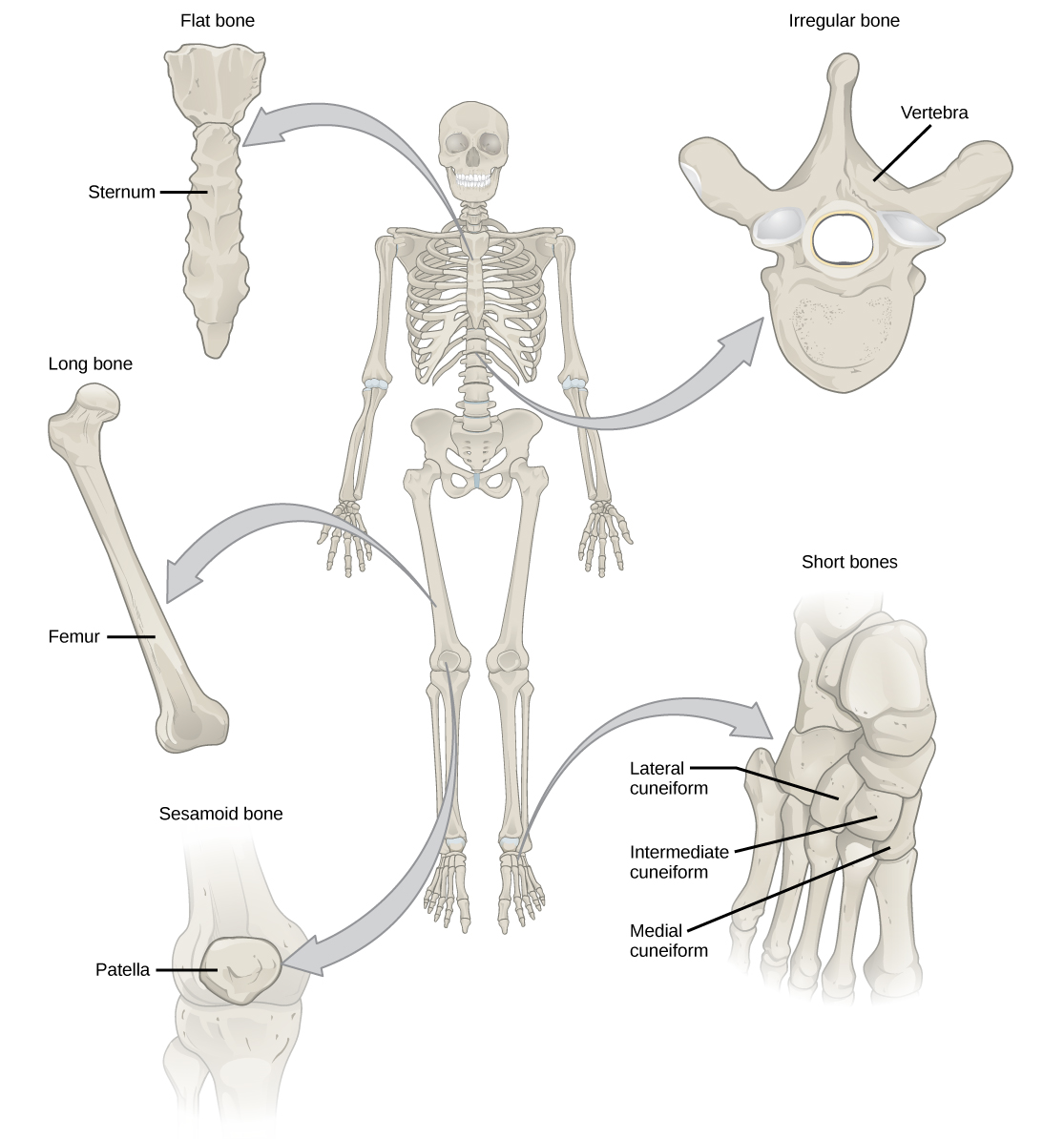
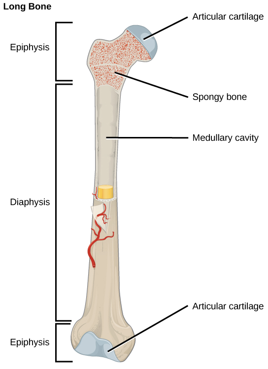
العظام الطويلة أطول من اتساعها ولها عمود وطرفان. يحتوي الحجاب الحاجز، أو العمود المركزي، على نخاع العظم في تجويف النخاع. تُغطى الأطراف المستديرة، المشاشية، بغضاريف مفصلية ومليئة بنخاع العظم الأحمر الذي ينتج خلايا الدم (الشكل\(\PageIndex{2}\)). معظم عظام الأطراف هي عظام طويلة - على سبيل المثال، عظم الفخذ والساق والزند والكعبرة. وتشمل الاستثناءات من ذلك الرضفة وعظام المعصم والكاحل.
العظام القصيرة، أو العظام المكعبة، هي عظام لها نفس العرض والطول، مما يمنحها شكلًا يشبه المكعب. على سبيل المثال، عظام الرسغ (الرسغ) والكاحل (الترسبات) هي عظام قصيرة (الشكل\(\PageIndex{1}\)).
العظام المسطحة هي عظام رقيقة وعريضة نسبيًا توجد في الأماكن التي تتطلب حماية واسعة للأعضاء أو عندما تكون الأسطح العريضة للتعلق العضلي مطلوبة. ومن أمثلة العظام المسطحة القص (عظم الثدي) والأضلاع والكتف (شفرات الكتف) وسقف الجمجمة (الشكل\(\PageIndex{1}\)).
العظام غير المنتظمة هي عظام ذات أشكال معقدة. قد تحتوي هذه العظام على أسطح قصيرة أو مسطحة أو مسننة أو متموجة. ومن أمثلة العظام غير المنتظمة الفقرات وعظام الورك والعديد من عظام الجمجمة.
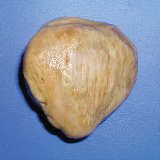
عظام السمسم هي عظام صغيرة ومسطحة وتشكلها بشكل مشابه لبذور السمسم. الرضفات هي عظام السمسم (الشكل\(\PageIndex{3}\)). تتطور عظام السمسم داخل الأوتار ويمكن العثور عليها بالقرب من المفاصل في الركبتين واليدين والقدمين.
العظام الجراحية هي عظام صغيرة ومسطحة وغير منتظمة الشكل. يمكن العثور عليها بين العظام المسطحة للجمجمة. وهي تختلف في العدد والشكل والحجم والموضع.
الأنسجة العظمية
تعتبر العظام أعضاء لأنها تحتوي على أنواع مختلفة من الأنسجة، مثل الدم والأنسجة الضامة والأعصاب وأنسجة العظام. تشكل الخلايا العظمية، وهي الخلايا الحية لأنسجة العظام، المصفوفة المعدنية للعظام. هناك نوعان من الأنسجة العظمية: المدمجة والإسفنجية.
أنسجة العظام المدمجة
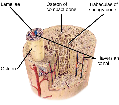
يشكل العظم المضغوط (أو العظم القشري) الطبقة الخارجية الصلبة لجميع العظام ويحيط بالتجويف النخاعي أو نخاع العظام. يوفر الحماية والقوة للعظام. يتكون النسيج العظمي المضغوط من وحدات تسمى العظام أو أنظمة Haversian. العظام عبارة عن هياكل أسطوانية تحتوي على مصفوفة معدنية وخلايا عظمية حية متصلة بالقناليكولي التي تنقل الدم. يتم محاذاتها بالتوازي مع المحور الطويل للعظام. تتكون كل عظمة من صفائح، وهي طبقات من المصفوفة المدمجة التي تحيط بقناة مركزية تسمى قناة هافرسيان. تحتوي قناة Haversian (القناة العظمية) على الأوعية الدموية للعظام والألياف العصبية (الشكل\(\PageIndex{4}\)). تتم محاذاة العظام في الأنسجة العظمية المضغوطة في نفس الاتجاه على طول خطوط الإجهاد وتساعد العظام على مقاومة الانحناء أو الكسر. لذلك، تكون الأنسجة العظمية المدمجة بارزة في مناطق العظام التي يتم فيها تطبيق الضغوط في اتجاهات قليلة فقط.
آرت كونيكشن
أي من العبارات التالية عن الأنسجة العظمية خاطئة؟
- يتكون النسيج العظمي المضغوط من عظام أسطوانية يتم محاذاتها بحيث تنتقل على طول العظم.
- تحتوي قنوات Haversian على الأوعية الدموية فقط.
- تحتوي قنوات Haversian على الأوعية الدموية والألياف العصبية.
- يوجد النسيج الإسفنجي في الجزء الداخلي من العظم، ويوجد نسيج عظمي مضغوط في الجزء الخارجي.
أنسجة العظام الإسفنجية
في حين أن الأنسجة العظمية المدمجة تشكل الطبقة الخارجية لجميع العظام، فإن العظم الإسفنجي أو العظم الإسفنجي يشكل الطبقة الداخلية لجميع العظام. لا تحتوي الأنسجة العظمية الإسفنجية على عظام تشكل نسيجًا عظميًا مضغوطًا. وبدلاً من ذلك، تتكون من الترابيكوليا، وهي عبارة عن صفائح مرتبة على شكل قضبان أو ألواح. تم العثور على نخاع العظم الأحمر بين التربيق. تقوم الأوعية الدموية داخل هذا النسيج بتوصيل العناصر الغذائية إلى الخلايا العظمية وإزالة النفايات. يشكل نخاع العظم الأحمر لعظم الفخذ والجزء الداخلي من العظام الكبيرة الأخرى، مثل الدقاق، خلايا الدم.
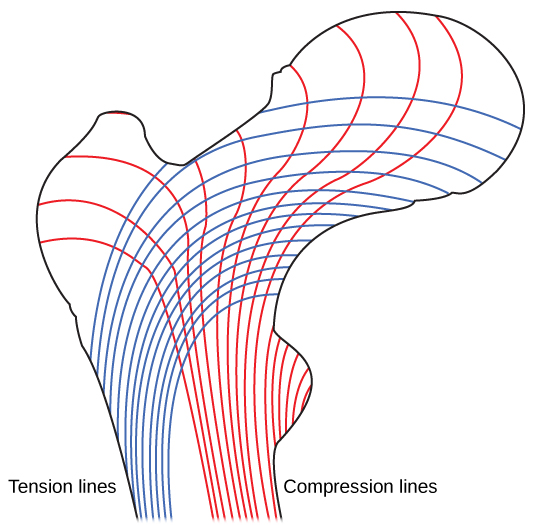
يقلل العظم الإسفنجي من كثافة العظام ويسمح لنهايات العظام الطويلة بالضغط نتيجة الضغوط المفروضة على العظام. تبرز العظام الإسفنجية في مناطق العظام التي لا تتعرض لضغوط شديدة أو حيث تأتي الضغوط من عدة اتجاهات. تتعرض مشاشات العظام، مثل عنق عظم الفخذ، للضغط من عدة اتجاهات. تخيل وضع صورة مؤطرة ثقيلة مسطحة على الأرض. يمكنك رفع جانب واحد من الصورة باستخدام عود أسنان إذا كان المسواك عموديًا على الأرض والصورة. الآن قم بحفر حفرة وألصق المسواك في الحائط لتعليق الصورة. في هذه الحالة، تتمثل وظيفة المسواك في نقل الضغط الهبوطي للصورة إلى الحائط. القوة الموجودة في الصورة تتجه مباشرة إلى الأرض، ولكن القوة التي تؤثر على المسواك هي سحب سلك الصورة لأسفل ودفع الجزء السفلي من الفتحة في الجدار لأعلى. سوف ينكسر المسواك مباشرة عند الحائط.
رقبة عظم الفخذ أفقية مثل المسواك في الحائط. إن وزن الجسم يدفعه إلى الأسفل بالقرب من المفصل، لكن الحجاب الحاجز الرأسي لعظم الفخذ يدفعه لأعلى عند الطرف الآخر. يجب أن تكون رقبة عظم الفخذ قوية بما يكفي لنقل القوة الهبوطية لوزن الجسم أفقيًا إلى العمود الرأسي لعظم الفخذ (الشكل\(\PageIndex{5}\)).

أنواع الخلايا في العظام
تتكون العظام من أربعة أنواع من الخلايا: بانيات العظم، الخلايا العظمية، الخلايا العظمية، وخلايا العظم. بانيات العظم هي خلايا العظام المسؤولة عن تكوين العظام. تقوم بانيات العظم بتجميع وإفراز الجزء العضوي والجزء غير العضوي من المصفوفة خارج الخلية لأنسجة العظام وألياف الكولاجين. تصبح بانيات العظم محاصرة في هذه الإفرازات وتتفرق إلى خلايا عظمية أقل نشاطًا. الخلايا الآكلة للعظام هي خلايا عظمية كبيرة تصل إلى 50 نواة. إنها تزيل بنية العظام عن طريق إطلاق الإنزيمات والأحماض الليزوزومية التي تعمل على إذابة المصفوفة العظمية. تساعد هذه المعادن، المنبعثة من العظام إلى الدم، على تنظيم تركيزات الكالسيوم في سوائل الجسم. يمكن أيضًا إعادة امتصاص العظام لإعادة تشكيلها، إذا تغيرت الضغوط المطبقة. الخلايا العظمية هي خلايا عظمية ناضجة وهي الخلايا الرئيسية في النسيج الضام العظمي؛ لا يمكن أن تنقسم هذه الخلايا. تحافظ الخلايا العظمية على بنية العظام الطبيعية عن طريق إعادة تدوير الأملاح المعدنية في المصفوفة العظمية. الخلايا العظمية هي خلايا جذعية حرشفية تنقسم لإنتاج خلايا ابنة تتمايز إلى بانيات عظمية. خلايا هشاشة العظام مهمة في إصلاح الكسور.
تطوير العظام
التعظم، أو تكوين العظم، هو عملية تكوين العظام بواسطة بانيات العظم. يختلف التعظم عن عملية التكلس؛ بينما يحدث التكلس أثناء تعظم العظام، يمكن أن يحدث أيضًا في الأنسجة الأخرى. يبدأ التعظم بعد حوالي ستة أسابيع من الإخصاب في الجنين. قبل هذا الوقت، كان الهيكل العظمي الجنيني يتكون بالكامل من الأغشية الليفية والغضاريف الهيالينية. يسمى تطور العظام من الأغشية الليفية بالتحجر الغشائي؛ ويسمى التطور من الغضروف الهياليني بالتحجر الداخلي. يستمر نمو العظام حتى سن 25 تقريبًا. يمكن أن تنمو العظام في السماكة طوال الحياة، ولكن بعد سن 25، يعمل التعظم بشكل أساسي في إعادة تشكيل العظام وإصلاحها.
التحجر داخل الغشاء
التعظم داخل الغشاء هو عملية تطور العظام من الأغشية الليفية. وتشارك في تكوين العظام المسطحة للجمجمة والفك السفلي والترقوة. يبدأ التعظم عندما تشكل الخلايا الوسيطة نموذجًا للعظم المستقبلي. ثم تتمايز إلى بانيات عظمية في مركز التحجر. تفرز الخلايا العظمية المصفوفة خارج الخلية وترسب الكالسيوم، مما يقوي المصفوفة. يستمر الجزء غير المعدني من العظم أو العظم في التكون حول الأوعية الدموية، مكونًا العظام الإسفنجية. يتحول النسيج الضام في المصفوفة إلى نخاع عظمي أحمر في الجنين. يتم إعادة تشكيل العظم الإسفنجي إلى طبقة رقيقة من العظام المدمجة على سطح العظم الإسفنجي.
التحجر الداخلي
التعظم الداخلي هو عملية تطور العظام من الغضروف الهياليني. تتشكل جميع عظام الجسم، باستثناء العظام المسطحة للجمجمة والفك السفلي والترقوة، من خلال التعظم الداخلي.
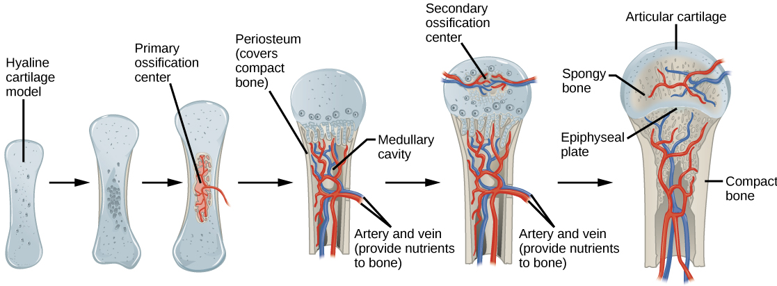
في العظام الطويلة، تشكل الخلايا الغضروفية نموذجًا لغضاريف الهيالين. عند الاستجابة للإشارات التنموية المعقدة، تبدأ المصفوفة في التكلس. يمنع هذا التكلس انتشار العناصر الغذائية في المصفوفة، مما يؤدي إلى موت الخلايا الغضروفية وفتح تجاويف في غضروف الحجاب الحاجز. تغزو الأوعية الدموية التجاويف، وتقوم بانيات العظم وخلايا العظم بتعديل مصفوفة الغضروف المتكلس إلى عظم إسفنجي. تقوم الخلايا العظمية بعد ذلك بتكسير بعض العظام الإسفنجية لإنشاء تجويف نخاعي أو نخاعي في وسط الحجاب الحاجز. يشكل النسيج الضام الكثيف وغير المنتظم غمدًا (السمحاق) حول العظام. يساعد السمحاق في ربط العظام بالأنسجة والأوتار والأربطة المحيطة. يستمر العظم في النمو والاستطالة مع انقسام الخلايا الغضروفية في المشاشية.
في المرحلة الأخيرة من نمو العظام قبل الولادة، تبدأ مراكز المشاشية في التكلس. تتشكل مراكز التعظم الثانوية في المشاش حيث تدخل الأوعية الدموية وبانيات العظم إلى هذه المناطق وتحول الغضروف الهياليني إلى عظم إسفنجي. حتى سن المراهقة، يستمر الغضروف الهياليني في الصفيحة المشاشية (صفيحة النمو)، وهي المنطقة الواقعة بين الحجاب الحاجز والكتلة المسؤولة عن النمو الطولي للعظام الطويلة (الشكل 1)\(\PageIndex{6}\)).
Growth of Bone
Long bones continue to lengthen, potentially until adolescence, through the addition of bone tissue at the epiphyseal plate. They also increase in width through appositional growth.
Lengthening of Long Bones
Chondrocytes on the epiphyseal side of the epiphyseal plate divide; one cell remains undifferentiated near the epiphysis, and one cell moves toward the diaphysis. The cells, which are pushed from the epiphysis, mature and are destroyed by calcification. This process replaces cartilage with bone on the diaphyseal side of the plate, resulting in a lengthening of the bone.
Long bones stop growing at around the age of 18 in females and the age of 21 in males in a process called epiphyseal plate closure. During this process, cartilage cells stop dividing and all of the cartilage is replaced by bone. The epiphyseal plate fades, leaving a structure called the epiphyseal line or epiphyseal remnant, and the epiphysis and diaphysis fuse.
Thickening of Long Bones
Appositional growth is the increase in the diameter of bones by the addition of bony tissue at the surface of bones. Osteoblasts at the bone surface secrete bone matrix, and osteoclasts on the inner surface break down bone. The osteoblasts differentiate into osteocytes. A balance between these two processes allows the bone to thicken without becoming too heavy.
Bone Remodeling and Repair
Bone renewal continues after birth into adulthood. Bone remodeling is the replacement of old bone tissue by new bone tissue. It involves the processes of bone deposition by osteoblasts and bone resorption by osteoclasts. Normal bone growth requires vitamins D, C, and A, plus minerals such as calcium, phosphorous, and magnesium. Hormones such as parathyroid hormone, growth hormone, and calcitonin are also required for proper bone growth and maintenance.
Bone turnover rates are quite high, with five to seven percent of bone mass being recycled every week. Differences in turnover rate exist in different areas of the skeleton and in different areas of a bone. For example, the bone in the head of the femur may be fully replaced every six months, whereas the bone along the shaft is altered much more slowly.
Bone remodeling allows bones to adapt to stresses by becoming thicker and stronger when subjected to stress. Bones that are not subject to normal stress, for example when a limb is in a cast, will begin to lose mass. A fractured or broken bone undergoes repair through four stages:
- Blood vessels in the broken bone tear and hemorrhage, resulting in the formation of clotted blood, or a hematoma, at the site of the break. The severed blood vessels at the broken ends of the bone are sealed by the clotting process, and bone cells that are deprived of nutrients begin to die.
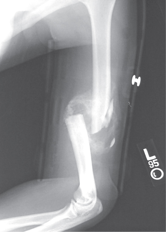
- Within days of the fracture, capillaries grow into the hematoma, and phagocytic cells begin to clear away the dead cells. Though fragments of the blood clot may remain, fibroblasts and osteoblasts enter the area and begin to reform bone. Fibroblasts produce collagen fibers that connect the broken bone ends, and osteoblasts start to form spongy bone. The repair tissue between the broken bone ends is called the fibrocartilaginous callus, as it is composed of both hyaline and fibrocartilage (Figure \(\PageIndex{7}\)). Some bone spicules may also appear at this point.
- The fibrocartilaginous callus is converted into a bony callus of spongy bone. It takes about two months for the broken bone ends to be firmly joined together after the fracture. This is similar to the endochondral formation of bone, as cartilage becomes ossified; osteoblasts, osteoclasts, and bone matrix are present.
- The bony callus is then remodelled by osteoclasts and osteoblasts, with excess material on the exterior of the bone and within the medullary cavity being removed. Compact bone is added to create bone tissue that is similar to the original, unbroken bone. This remodeling can take many months, and the bone may remain uneven for years.
Scientific Method Connection: Decalcification of Bones
Question: What effect does the removal of calcium and collagen have on bone structure?
Background: Conduct a literature search on the role of calcium and collagen in maintaining bone structure. Conduct a literature search on diseases in which bone structure is compromised.
Hypothesis: Develop a hypothesis that states predictions of the flexibility, strength, and mass of bones that have had the calcium and collagen components removed. Develop a hypothesis regarding the attempt to add calcium back to decalcified bones.
Test the hypothesis: Test the prediction by removing calcium from chicken bones by placing them in a jar of vinegar for seven days. Test the hypothesis regarding adding calcium back to decalcified bone by placing the decalcified chicken bones into a jar of water with calcium supplements added. Test the prediction by denaturing the collagen from the bones by baking them at 250°C for three hours.
Analyze the data: Create a table showing the changes in bone flexibility, strength, and mass in the three different environments.
Report the results: Under which conditions was the bone most flexible? Under which conditions was the bone the strongest?
Draw a conclusion: Did the results support or refute the hypothesis? How do the results observed in this experiment correspond to diseases that destroy bone tissue?
Summary
Bone, or osseous tissue, is connective tissue that includes specialized cells, mineral salts, and collagen fibers. The human skeleton can be divided into long bones, short bones, flat bones, and irregular bones. Compact bone tissue is composed of osteons and forms the external layer of all bones. Spongy bone tissue is composed of trabeculae and forms the inner part of all bones. Four types of cells compose bony tissue: osteocytes, osteoclasts, osteoprogenitor cells, and osteoblasts. Ossification is the process of bone formation by osteoblasts. Intramembranous ossification is the process of bone development from fibrous membranes. Endochondral ossification is the process of bone development from hyaline cartilage. Long bones lengthen as chondrocytes divide and secrete hyaline cartilage. Osteoblasts replace cartilage with bone. Appositional growth is the increase in the diameter of bones by the addition of bone tissue at the surface of bones. Bone remodeling involves the processes of bone deposition by osteoblasts and bone resorption by osteoclasts. Bone repair occurs in four stages and can take several months.
Art Exercise
Figure \(\PageIndex{4}\): Which of the following statements about bone tissue is false?
- Compact bone tissue is made of cylindrical osteons that are aligned such that they travel the length of the bone.
- Haversian canals contain blood vessels only.
- Haversian canals contain blood vessels and nerve fibers.
- Spongy tissue is found on the interior of the bone, and compact bone tissue is found on the exterior.
- Answer
-
B
Glossary
- appositional growth
- increase in the diameter of bones by the addition of bone tissue at the surface of bones
- bone
- (also, osseous tissue) connective tissue that constitutes the endoskeleton
- bone remodeling
- replacement of old bone tissue by new bone tissue
- calcification
- process of deposition of mineral salts in the collagen fiber matrix that crystallizes and hardens the tissue
- compact bone
- forms the hard external layer of all bones
- diaphysis
- central shaft of bone, contains bone marrow in a marrow cavity
- endochondral ossification
- process of bone development from hyaline cartilage
- epiphyseal plate
- region between the diaphysis and epiphysis that is responsible for the lengthwise growth of long bones
- epiphysis
- rounded end of bone, covered with articular cartilage and filled with red bone marrow, which produces blood cells
- flat bone
- thin and relatively broad bone found where extensive protection of organs is required or where broad surfaces of muscle attachment are required
- Haversian canal
- contains the bone’s blood vessels and nerve fibers
- intramembranous ossification
- process of bone development from fibrous membranes
- irregular bone
- bone with complex shapes; examples include vertebrae and hip bones
- lamella
- layer of compact tissue that surrounds a central canal called the Haversian canal
- long bone
- bone that is longer than wide, and has a shaft and two ends
- osteoblast
- bone cell responsible for bone formation
- osteoclast
- large bone cells with up to 50 nuclei, responsible for bone remodeling
- osteocyte
- mature bone cells and the main cell in bone tissue
- osseous tissue
- connective tissue that constitutes the endoskeleton
- ossification
- (also, osteogenesis) process of bone formation by osteoblasts
- osteon
- cylindrical structure aligned parallel to the long axis of the bone
- resorption
- process by which osteoclasts release minerals stored in bones
- sesamoid bone
- small, flat bone shaped like a sesame seed; develops inside tendons
- short bone
- bone that has the same width and length, giving it a cube-like shape
- spongy bone tissue
- forms the inner layer of all bones
- suture bone
- small, flat, irregularly shaped bone that forms between the flat bones of the cranium
- trabeculae
- lamellae that are arranged as rods or plates